ISC 11>Content>Unit-4>Biomolecules :Nucleic acid
Structure and functions of DNA, types of RNA. Differences between DNA and RNA
|
NUCLEOTIDES: (BUILDING BLOCKS OF NUCLEIC ACID)
COMPOSITION:
- Nucleotides contain carbon, hydrogen, oxygen, nitrogen and phosphorus.
- Each nucleotide is made up of following components.
- Ribose sugar
- Phosphoric acid
- Nitrogenous bases
|
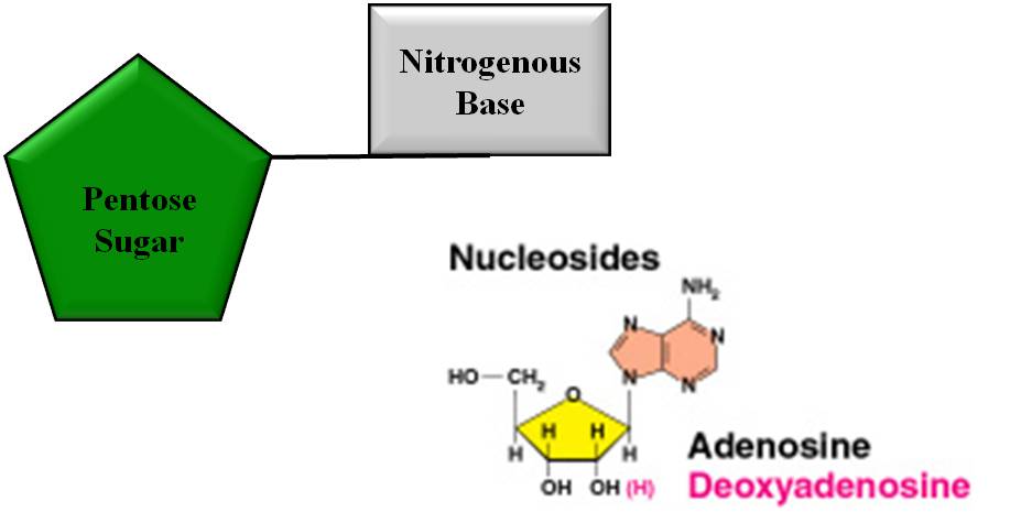
1. Ribose sugar
The sugar molecule in the nucleotide is a 5-carbon pentose sugar. It is represented by either ribose sugar (C5H10O5) or deoxyribose sugar (C5H10O4). 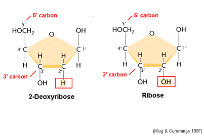
|
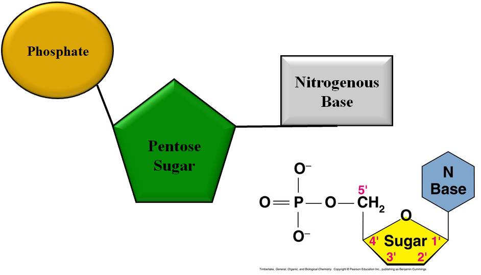
2. Phosphoric acid
It is a made up of three acid groups.
These acid group react with OH group of ribose or deoxyribose to form pentose monophosphate esters. 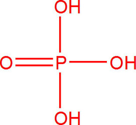
|
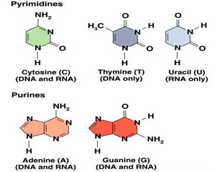
3. Nitrogenous bases:
- Nitrogenous bases are in the form of heterocyclic aromatic rings, formed of carbon and nitrogen.
- Two types of nitrogen bases occur, namely
- Purines, which have a double ring structure.(Adenine and Guanine)
- Pyrimidines, which have a single ring structure. (Cytosine, Thymine and Uracil)
|
|
FORMATION OF NUCLEOSIDES & NUCLEOTIDES
|
NUCLEOSIDE:
|
NUCLEOTIDE:
|
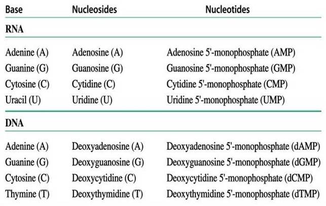
FUNCTIONS OF NUCLEOTIDES
- Building blocks of nucleic acid.(RNA, DNA)
- Regulatory chemicals (cAMP)
- Formation of energy carriers (ADP and ATP)
- Formation of coenzymes (NAD, NADP, FMN, FAD)
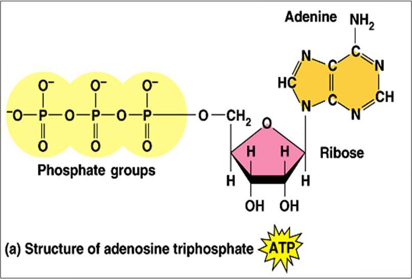
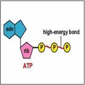
HIGHER NUCLEOTIDES : (The energy carriers)
- Nucleotides with more than one phosphate molecule is termed higher nucleotides.
- Examples: ADP, ATP, GDP, GTP, CDP, CTP,
TDP, TTP, UDP and UTP.
- ADP - Adenosine diphosphate
- ATP – Adenosine triphosphate
- CDP -Cytidine diphosphate
- CTP – Cytidine triphosphate
- TDP - Thymidine diphosphate
- TTP – Thymidine triphosphate
- GDP- Guanosine diphosphate
- GTP – Guanosine triphosphate
- UDP -Uridine diphosphate
- UTP –Uridine triphosphate
High energy or energy rich bond
|
|
|
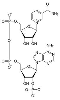
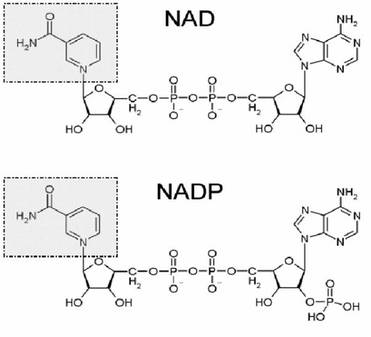
COENZYMES
- Organic carrier molecule cooperating with an enzyme in promoting a chemical reaction in a cell.
- Composition: Formed from nucleotides by replacing nitrogenous base with an vitamin.
- Mode of action: In many reaction, removal of group of atoms and addition to other. This is done with enzyme and coenzyme helps in its transfer to another acceptor compound.
- Examples:
- NAD: Nicotinamide adenine dinucleotide
- NADP : Nicotinamide adenine dinucleotide phosphate
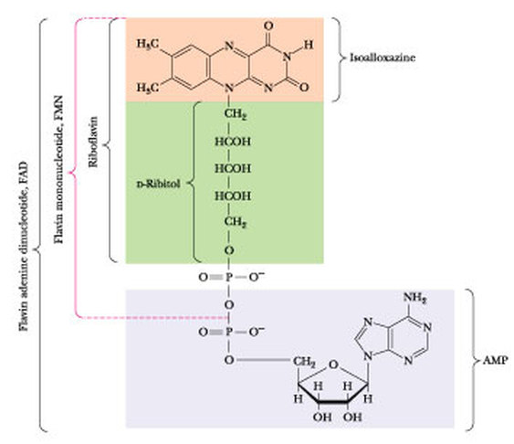
- FMN : Flavin mononucleotide
- FAD : Flavin adenine dinucleotide
- Coenzyme A
|
NAD :
The compound is a dinucleotide, since it consists of two nucleotides joined through their phosphate groups, with one nucleotide containing an adenine base and the other containing nicotinamide. |
|
|
|
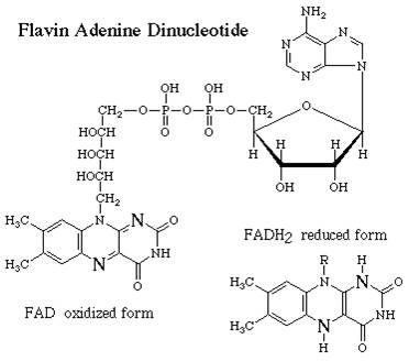
FAD
This compound is made up of one nucleotide containing ribose and adenine and one with an unusual structure involving a linear molecule RIBITOL instead of ribose. |
|
|
NUCLEIC ACID
HISTORY OF NUCLEIC ACID
|
1. Friedrich Miescher (1844-1895)
In 1869, Friedrich Miescher isolated "nuclein," DNA with associated proteins, from cell nuclei. He was the first to identify DNA as a distinct molecule. 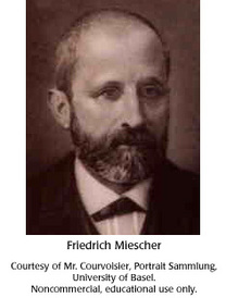
|
2. Albrecht Kossel (1853 – 1927)
Kossel isolated and described the five organic compounds that are present in nucleic acid adenine, cytosine, guanine, thymine, and uracil. These compounds were later shown to be nucleobases. 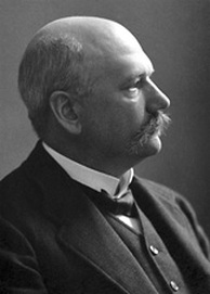
|
3. Oscar Hertwig (1849– 1922)
He discovered fertilization of sea urchins, he recognized the role of the cell nucleus during inheritance and chromosome reduction during meiosis 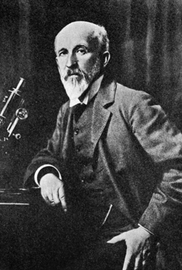
|
4. Richard Altman ( (1852 – 1900)) :Altmann is credited for coining the term "nucleic acid", in exchange for Friedrich Miescher's (1844-1895) nuclein, when it was demonstrated that nuclein had acidic properties.
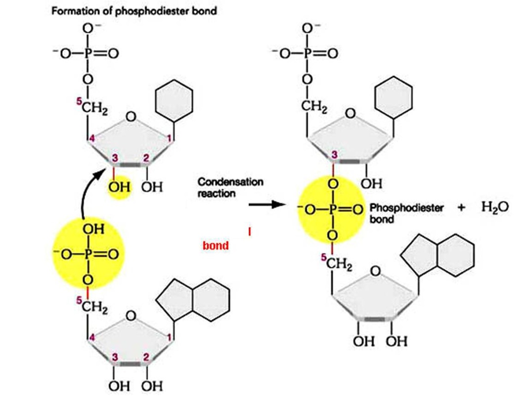
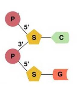
FORMATION OF DINUCLEOTIDE AND POLYNUCLEOTIDE
|
|
|
|
NUCLEIC ACID
- Nucleic acids are a polynucleotides consisting of repeated units of mononucleotides.
- Classification : Nucleic acids are classified into
- DNA : DEOXYRIBONUCLEIC ACID
- RNA : RIBONUCLEIC ACID
DNA: HISTORY
1. PHOEBUS LEVENE: Phoebus Levene (1869-1940) had discovered that the cell nucleus contains two types of nucleic acids - DNA and RNA. He also found that DNA contained the four bases, deoxyribose, and a phosphate group.
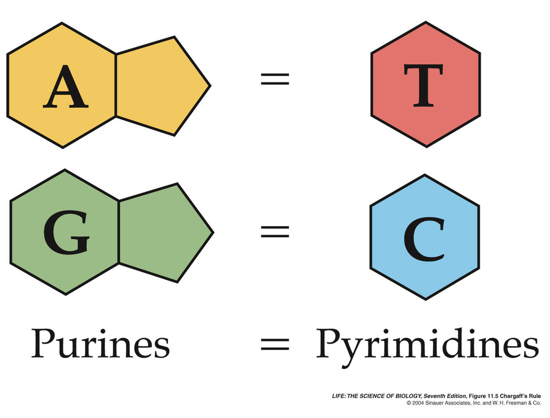
2. ERWIN CHARGAFF
In 1950, Erwin Chargaff published a paper stating that in DNA of any given species, the ratio of adenine to thymine is equal, as is the ratio of cytosine to guanine. This is known as Chargaff's ratios and it was a crucial clue that helped solve the structure of DNA. Chargaff's ratios are universal: all forms of life obey this rule. Only the balance of A-T pairs and C-G pairs varies between species.
|
|
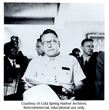
3. ROSALIND FRANKLIN
Rosalind Franklin and Raymond Gosling obtained this X-ray diffraction pattern, which triggered the idea that DNA was a helix.
She produced the X-ray crystallography pictures of DNA which Watson and Crick used to determine the structure of double-stranded DNA.
Biography : Rosalind Elsie Franklin (1920-1958)
She produced the X-ray crystallography pictures of DNA which Watson and Crick used to determine the structure of double-stranded DNA.
Biography : Rosalind Elsie Franklin (1920-1958)
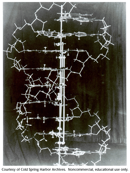
4. WATSON AND CRICK
James D. Watson and Francis Crick were the two co-discoverers of the structure of DNA in 1953. They used x-ray diffraction data collected by Rosalind Franklin and proposed the double helix or spiral staircase structure of the DNA molecule.
WATSON AND CRICK MODEL OF DNA
|
The six feet tall metal DNA model made by Watson and Crick in 1953.
|
Discovering the double helix structure of DNA, James Watson, video with 3D animation and narration
|
|
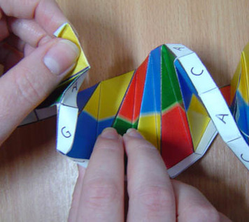
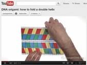
ACTIVITY
| ||||
In 1953, J.D. Watson and F.H.C. Crick proposed a precise three dimensional model of DNA structure based on model building studies,
base composition and X-ray diffraction studies. This model is popularly known as the DNA double helix.
base composition and X-ray diffraction studies. This model is popularly known as the DNA double helix.
|
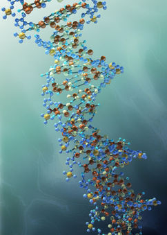
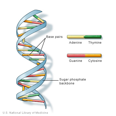
DNA DOUBLE HELIX
|
SCHEMATIC REPRESENTATION OF DNA DOUBLE HELIX
|
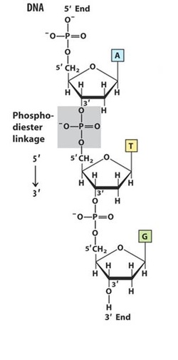
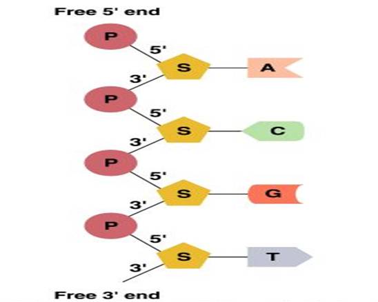
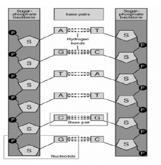
- Two polynuleotide chains are coiled around a central axis in the form of a right handed double helix.
- The backbone of each strand consists of alternating sugar and phosphate. The bases project inwards and they are perpendicular to the central axis.
- The 2 strands run in opposite direction (ie.) they are antiparallel. Each polynucleotide chain has 5’ phosphorylated and 3’ hydroxyl ends
- The strands are complementary to each other due to specific base pairing of the opposite strands. Base pairing occurs through hydrogen bonding and it is specific. Adenine pairs with thymine through two hydrogen bonds. Guanine pairs with cytosine with three hydrogen bonds.
- Major and minor grooves are present on the double helix.
- One turn of helix measures 34 angstrom and has 10 nucleotides.
- Distance between adjacent nucleotides is 3.4 angstrom.
- The diameter of the helix is 20 angstrom.
FUNCTIONS OF DNA
- Carries hereditary information.
- Controls the metabolic activity of the cell.
- Enables cell to maintain, grow and divide.
- Contributes to variation by undergoing mutation.
- Brings about differentiation of cells
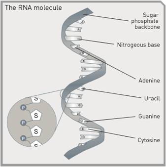
RNA : RIBONUCLEIC ACID
|
|
TYPES OF RNA
- There are mainly three types of RNAs in all prokaryotic and eukaryotic cells.
- They are
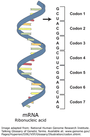
- Messenger RNA (mRNA)
- Transfer RNA (tRNA)
- Ribosomal RNA (rRNA).
- They differ from each other by size, formation and stability.
|
2. Ribosomal RNA (r RNA)

|
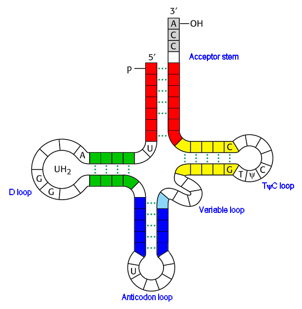
3. Transfer RNA (t RNA)
|
FUNCTIONS OF RNA
- Protein synthesis.
- Genetic material.
- Component of ribosomes.
- Associated with DNA replication.
- Some RNA have enzymatic activity (ribonuclease)
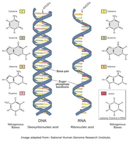
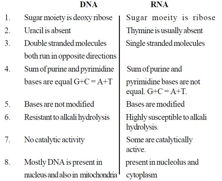
DIFFERENCES BETWEEN DNA & RNA
|
|
Why is DNA called an acid, especially since it contains a ‘base’ and doesn’t appear to have any dissociating hydrogen?
