ISC 12>CONTENT>STRUCTURE AND FUNCTION OF ANIMALS>1. DIGESTION AND ABSORPTION
SCOPE OF SYLLABUS
Calorific value of carbohydrates, proteins and fats per gram;
Structure and functions of the digestive organs and their associated glands,
Types of dentition (thecodont, heterodont, diphyodont) and dental formula of human;
Diagram of the digestive system with correct position of the organs and the associated glands;
Diagrammatic representation of T.S. of gut showing the four layers - histology of individual organs not required;
Physiology of digestion and absorption of food;
definition of bolus, peristalsis, deglutition, emulsification; assimilation of digested food;
disorders of the digestive system – Protein Energy Malnutrition ( PEM), indigestion, constipation, vomiting, jaundice,diarrhoea.
NUTRITIONPROCESS OF OBTAINING FOOD BY LIVING BEINGS, ITS DIGESTION AND UTILIZATION FOR ENERGY.
|
|
|
Watch the video and answer the following questions-
|
|
THE HUMAN DIGESTIVE SYSTEM
Diagram of the digestive system with correct position of the organs and the associated glands;
HOW TO DRAW THE DIAGRAM |
THE DIAGRAM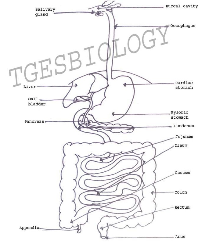
|
PARTS OF THE DIGESTIVE SYSTEM AND THEIR FUNCTIONS
Structure and functions of the digestive organs and their associated glands,
1. MOUTH AND BUCCAL CAVITY
|
The mouth is a transverse slit like opening bounded by two soft movable lips.
Buccal cavity is bound by- 1. Above- Palate
3. Sides- jaws Inside the buccal cavity is present a muscular tongue and teeth arranged on the jaws. |

|
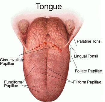
TONGUE
|
Is a skeletal muscle, helps to moves food in mouth, and to taste.
Taste buds- have papillae
|
|
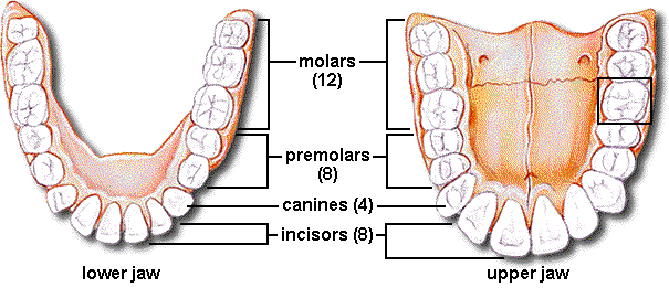
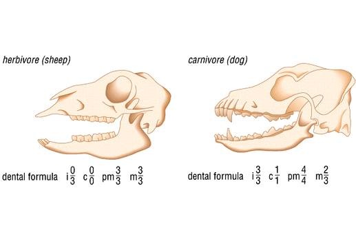
Types of dentition (thecodont, heterodont, diphyodont) and dental formula of human;
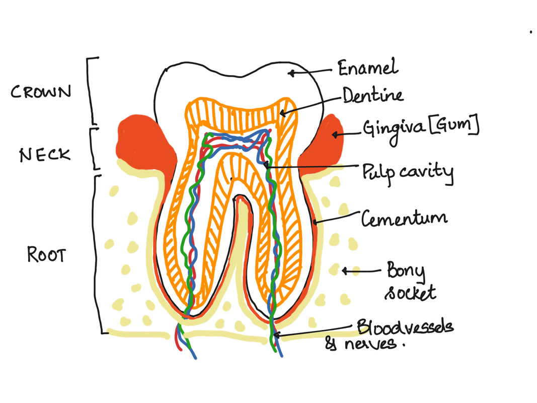
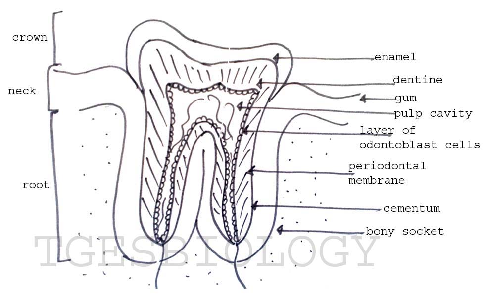
TEETH
http://www.student.loretto.org/humanbiology/BioLinks/chp5/media/c236f1_0.gif
FOUR types of teeth- incisors, canines, premolars, molars
The human teeth is
The human teeth is
- Heterodont: Humans have four types present. Incisors, canines, premolars and molars
- Thecodont: The neck and root are held in a bony socket.
- Diphyodont: There are two sets of teeth. The milk teeth are replaced by permanent teeth.
L.S.of molar tooth
|
Draw the diagram
|
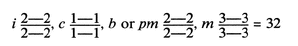
Competitive focus-Dental formula
|
Representation of the arrangement of human teeth.
http://www.merriam-webster.com/art/med/dental.gif
|
http://media.tiscali.co.uk/images/feeds/hutchinson/ency/c00764.jpg
|
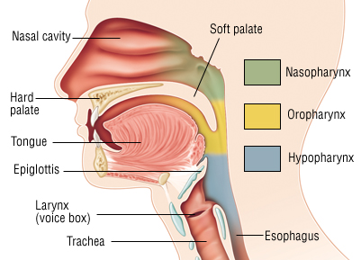
2. PHARYNX
|
It is a common passageway for air and food.
It participates in swallowing It has
It has three parts-
|
|
3. OESOPHAGUS
|
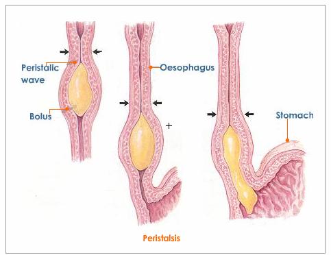
Oesophagus is a long tube starting from the pharynx to the stomach.
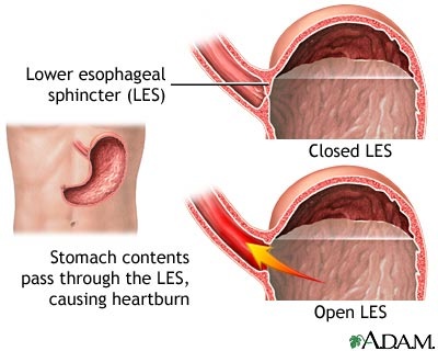
The opening of the oesophagus from the pharynx is called the gullet. It has a sphincter- upper oesophageal sphincter. The opening of the oesophagus to the stomach is guarded by lower oesophageal sphincter or the cardiac sphincter.
Peristalsis- contraction and relaxation of muscles which propagates in a wave down the muscular tube. |
|
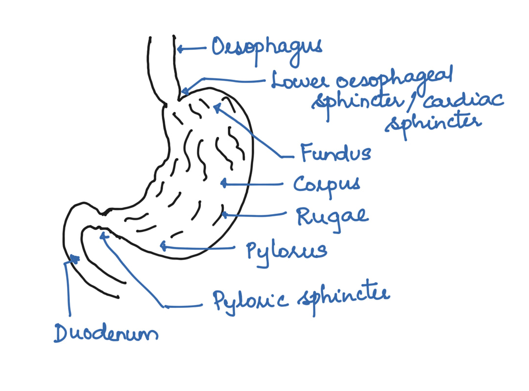
4. STOMACH
|
Large, muscular, somewhat J shaped sac, occupying the upper left part of the abdominal cavity.
Two regions- Cardiac and Pyloric. Shows the presence of longitudinal folds called rugae. Functions: Food storage, digestion, regulation of delivery. Produces gastric juice for digestion.
Gastrin: hormone secreted for stimulating digestion. |
|
Layers of the stomach wall
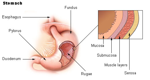
http://www.daviddarling.info/images/stomach.jpg
|
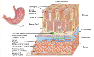
http://www.rivm.nl/interspeciesinfo/Images/stomach-figure-2_tcm75-26457.gif
|
What is heartburn or acidity?
Heartburn or pyrosisWhen the cardiac sphincter fails to close fully, the acidic chyme squeezes into the oesophagus.
This irritates the lining of oesophagus and causes heartburn. CARDIAC SPHINCTER It is less effective in infants hence regurgitation is more common among infants. |
|
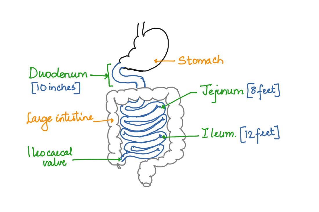
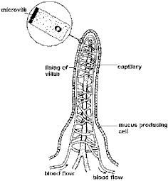
SMALL INTESTINE
|
Small intestine consists of
Ileum- absorption of digested food takes place here.
Functions of the small intestine:
Digestion: Neutralize acid from stomach, Add digestive enzymes and bile, breaks down proteins, carbohydrates and lipids to absorbable materials Absorption: 95% of food is absorbed here. Structure Mucosa adaptations: villi containing blood capillaries and lacteal. Cells of villi have small projections - microvilli. |
|
|
|
|
Villi in the small intestine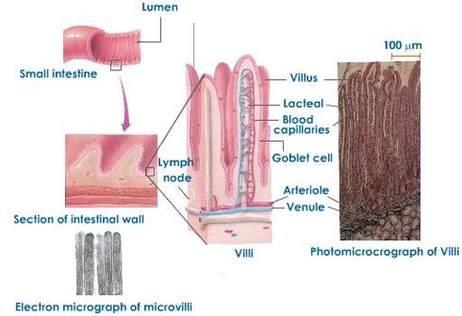
|
Small Intestine as principle site for absorption
|
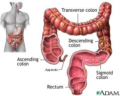
LARGE INTESTINE
Functions-
|
|
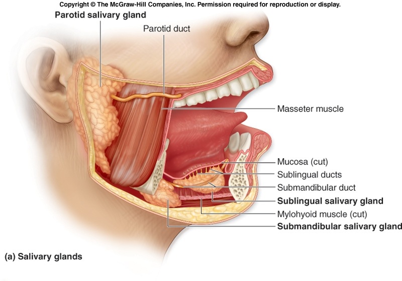
ASSOCIATED GLANDS
Salivary glands
|
Functions:
|
Gastric glands
|
Simple or branched tubular glands are present in the mucosa of the stomach.-
Gastric juice. Cardiac glands- secrete mucus Pyloric glands- secrete mucus Fundic glands four types of cells
|
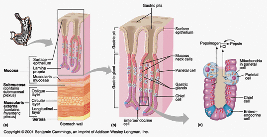
Gastric enzymes- pepsinogen, prorennin and gastric lipase
|
Intestinal glands
|
Microscopic glands present in the mucosa of small intestine.
Crypts of Lieberkuhn- multicellular, straight, tubular glands present throughout the mucosa. Secrete digestive enzyme and mucus. Brunner’s glands- compound tubular glands, found in the duodenum. Secrete alkaline watery juice. Intestinal secretion- the secretions from the intestinal glands is called succus entericus |
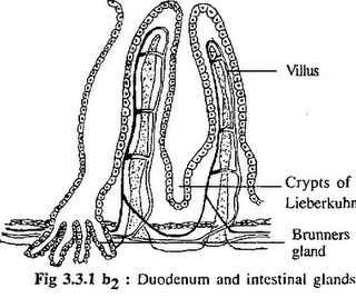
http://1.bp.blogspot.com/_qW0F6VNbvlM/TS09u0PKvaI/AAAAAAAAACY/RH1zN0JC10Y/s320/Intestinal+gland.png
|
Intestinal enzymes
Carbohydrate digesting enzymesIntestinal amylase
Maltase Sucrase Isomaltase Lactase Limit dextrase |
Protein digesting enzymesEnterokinase
Aminopeptidase Dipeptidase Tripeptidase Nuclease |
Fat digesting enzymesIntestinal lipase
|
Pancreas
|
Pancreas is a diffused leaf shaped gland.
It is formed of exocrine part and endocrine part. Exocrine part is made up of lobules or acini. Acinus is formed of glandular acinous cells. These cells secrete pancreatic juice. Enzymes in the pancreatic juice-
Two main type of cells are seen- Alpha cells- produces glucagon. Beta cells- produces insulin. |
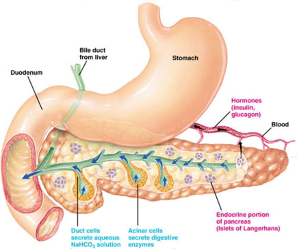
http://www.gopetsamerica.com/anatomy/illustrations/pancreas.jpg
|
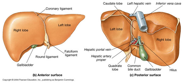
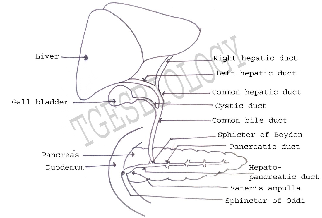
LIVER
|
Liver has two main lobes:- large right lobe and small left lobe.
In addition – quadrate lobe and caudate lobe are present.
The gallbladder is located beneath the right lobe of the liver. The primary function of the gallbladder is to store and concentrate bile. |
|
Liver - FunctionsThe primary functions of the liver are:
Lipogenesis- excess of glucose is converted to fats |
|
Glucose metabolism in the liver
|
Glycogenesis
Excess glucose in the blood is converted into glycogen and stored. |
Glycogenoglysis
When required glucose is obtained by hydrolysis of glycogen. |
Glyconeogenesis
Glucose is synthesized from amino acids or fatty acids and glycerol. |
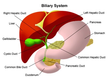
Bilary system
|
Bile is the greenish-yellow fluid (consisting of waste products, cholesterol, and bile salts) that is secreted by the liver cells to perform two primary functions:
|
Food by mastication is converted to bolus in the mouth.
It is passed to the stomach where churning and action of enzymes lead to formation of chyme, the partially digested food.
In the duodenum, complete digestion takes place forming the chyle.
It is passed to the stomach where churning and action of enzymes lead to formation of chyme, the partially digested food.
In the duodenum, complete digestion takes place forming the chyle.
Watch the videos showing the process of digestion.
|
|
|
