ISC 11>Content>Unit_1>Chapter_2_Kingdom Monera
SCOPE OF SYLLABUS
- Kingdom Monera: Bacteria - forms of bacteria, reproduction, gram +ve and gram –ve bacteria; economic importance- special emphasis on role of bacteria in sewage treatment, antibiotics, energy production; cyanobacteria: characteristic features; archaebacter(A brief idea of the role of different types of archaebacteria (methanogens, halophiles and thermoacidophils in their extreme environments),.
- Virus (characteristics features- link between living and non living, structure and name of the discoverers) and Viroid (definition only)
| moneraweb.docx |
CHARACTERISTICS OF KINGDOM MONERA
|
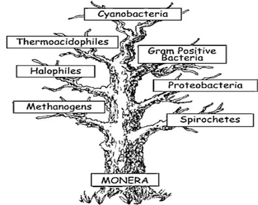
CLASSIFICATION OF MONERA
A) ON THE BASIS OF HABITATS (Evolutionary relationship)
|
|
OCCURRENCE AND DISTRIBUTION EVERY WHERE. WHY........?
- Extremely simple structure.
- Small size and consequent large surface-to- volume ratio. In order to maintain their small size, cell division occurs rapidly
- Resistance of vegetative cells to adverse environmental factors
- Formation of highly resistant endospores
- Diversity in their mode of nutrition
B) ON THE BASIS OF SHAPE
|
|
|
C) ON THE BASIS OF FLAGELLATION:
- All spirilla, about half of the bacilli and a small number of cocci are flagellated.
- Flagella vary both in number and arrangement according to two general patterns.
a. monotrichous – with a single flagellum
b. lophotrichous – with small bunches or tufts of flagella emerging from one end
c. amphitrichous – with flagella at both poles of the cell
2. In a peritrichous arrangement flagella are dispersed randomly over the surface of the cell.
3. Atrichous bacteria lack flagellum.
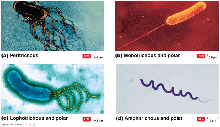
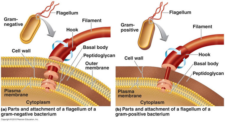
STRUCTURE OF FLAGELLA
D) ON THE BASIS OF MODE OF NUTRITION
Moneran organisms can be classified as either autotrophs or heterotrophs. Autotrophs are the producers of the world:
- Photoautotrophs: photosynthetic autotrophs (used to be called blue-green algae) that produce energy from light.
- Chemoautotrophs: produce energy from inorganic substances (e.g., Sulphur bacteria).
E) ON THE BASIS OF REACTIVITY WITH OXYGEN
Moneran organisms can be classified on the basis of their ability to react with oxygen:
- whether theymust react with oxygen to survive,
- whether they must be without oxygen to survive, or
- if they can survive with or without oxygen.
- There are three classes of oxygen reactivity:
- Obligate aerobes : Obligate aerobes require oxygen for respiration—they must have oxygen to grow
- Obligate anaerobes : must avoid oxygen like the plague—oxygen is a poison to them;
- Facultative anaerobes : Facultative anaerobes, which are happy to use O2 when available, but can survive without it.
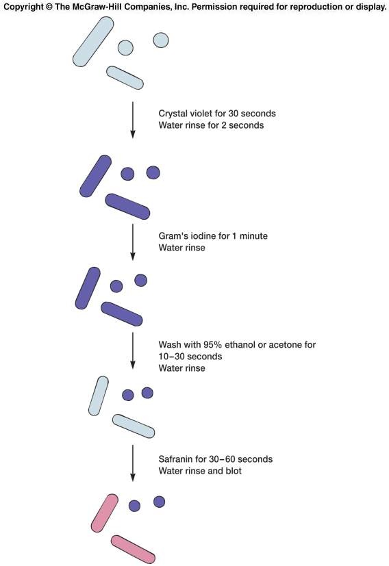
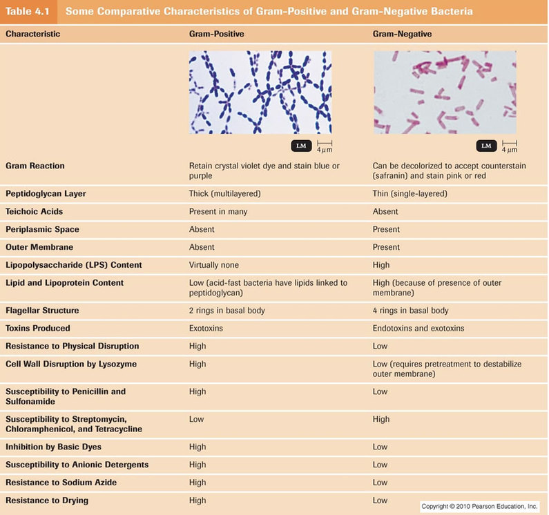
F) ON THE BASIS OF GRAM STAINING PROCESS
|
|
|
GRAM STAINING PROCESS : VIDEO
|
|
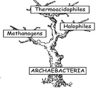
ARCHAEA
FEATURES OF ARCHAEBACTERIA
- Archaebacteria have no peptidoglycan in their cell walls
- The cell wall is made up of glycoproteins and polysaccarides.
- The cell wall envelopes have a high resistance to antibiotics and lytic agents due to difference in cell wall composition.
- They have a very different lipid bilayer making up the cell membranes
- The RNA polymerase of archae is very similar to that of eukaryotes
- The eukaryotes and archea ribosomal proteins are similar to each other
- Archaebacteria are about 1/10th of a micrometer to about 15 micrometer in size.
- A few are flagellated and the flagella structure is different from the flagella of other bacteria.
- The archaebacteria are non-pathogenic bateria that live in and around other organisms.
- Archaebacteria are autotrophs and use CO2 in atmosphere as a source of carbon for a process called carbon fixation.
HABITAT
Based on the nature of their habitats the archae are grouped into three , these are
- Methanogens
- Halophiles
- Thermoacidophiles.
- Methanogens:- They can reduce carbon dioxide into methane.They can only survive in the absence of oxygen.They produce marsh gas that one can observe as bubbles in stagnant waters. They are also present in the gut of cattle and termites. These bacteria are rod shaped or spherical and can be gram positive as well as negative.eg Methanobacterium, Methanococcus,Methanomicrobium
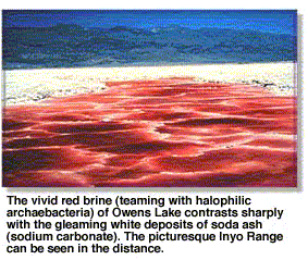
- Halophiles :- They are found in areas with very high salt concentrations sea. They contain bacteriorhodopsin, a red or orange pigment.eg Halobacterium,Halococcus, Natronabacterium
- Thermoacidiophiles :- Organisms that can survive in extremely high temperatures and low pH. They can survive at 100° Celsius with a pH of 2. Most of these organisms are anaerobic nature e.g Pyyrodictium, Pyrococcus, Sulfolobus, thermococcus, thermoproteus.
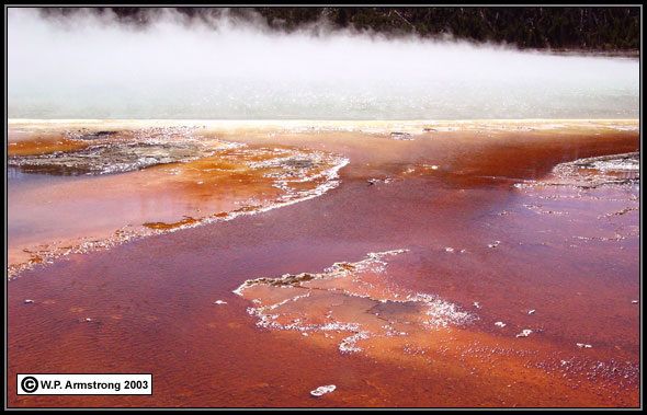
Bacteria Of Boiling Hot Springs In Yellowstone National Park
|
|
Methanogens
|
MORPHOLOGY
- Archaebacteria cells have diameters which range from about 0.0002–0.0004 in (0.5–1.0 micrometer). The volume of their cells is only around one-thousandth that of a typical eukaryotic cell. They have three main forms or shapes
- Spherical cells called cocci,
- Rod shaped cells called bacilli,
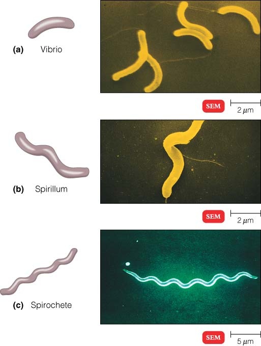
- Spiral shaped cells which can either be vibrio, spirillum , or spirochete
- Some other shapes may occur such as irregularly shaped lobed cells , needle-like filaments, almost perfectly rectangular rods , flat, square archaea, and filaments form aggregates or filaments and multicell colony.
STRUCTURE
- Archaebacteria, like all prokaryotes, have no membrane bound organelles.
- This means that the archaebacteria are without nuclei, mitochondria, endoplasmic reticula, lysosomes, Golgi complexes, or chloroplasts.
- The cells contain a thick cytoplasm that contains all of the molecules and compounds of metabolism and nutrition .
- Archaebacteria have a cell wall that contains no peptidoglycan. This rigid cell wall supports the cell, allowing an archaebacterium to maintain its shape, and protecting the cell from bursting when in a hypotonic environment.
- The DNA is a single circular molecule. This molecule is tightly wound and compact, and if stretched out would be more than 1,000 times longer than the actual cell. Little or no protein is associated with the DNA.
- Plasmids may be present in the archaebacterial cell. These are small, circular pieces of DNA that can duplicate independent of the larger, genomic DNA circle.
- Plasmids often code for particular enzymes or for antibiotic resistance.
ECONOMIC IMPORTANCE OF ARCHAEBACTERIA
- Methanogen archaebacteria are used in the generation of gobar gas from dung and sewage .
- Heat resistant thermophilic enzymes and restriction enzymes in biotechnology.
- Archaebacteria are used as biosensors.
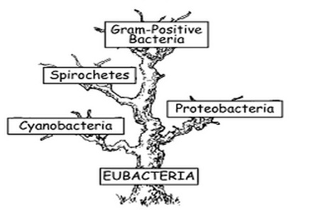
EUBACTERIA
HABITAT
The Eubacteria are cosmopolitan, they live everywhere, soils, water, faeces, decaying substances, bodies of plants and animals.
CLASSIFICATION
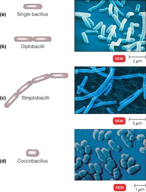
- There are three main criteria used to classify the Eubacteria, they are shape
- It is easy to distinguish the Eubacteria based on their shape. Bacterial cells have three main shapes:
- Cocci: round
- Bacilli: rod-shaped
- Spirilli: spiral-shaped
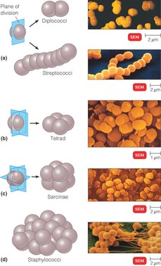
- Furthermore, bacteria can be classified by their growth characteristic patterns (Groupings). The prefix diplo- means that the cells are arranged in pairs. The prefix staphylo- means that the bacterial cells are arranged in clusters like grapes. The prefix strepto- means that the bacteria are arranged in a chain.
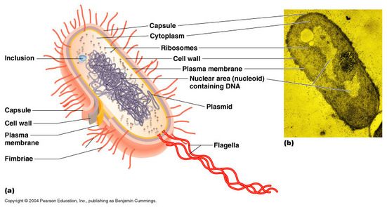
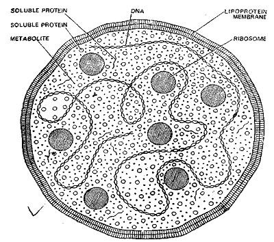
STRUCTURE OF BACTERIAL CELL
|
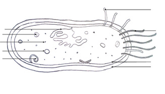
Copy and label : A bacterium cell
|
STRUCTURE
A) Bacterial Cell Wall
- The cell wall is a rigid structure that lies just outside the plasma membrane; it provides the characteristic shapes of the various procaryotes and protects them from osmotic lysis
- The cell walls of most bacteria contain peptidoglycan; the cell walls of archaea lack peptidoglycan and instead are composed of proteins, glycoptoteins, or polysaccharides
- The cell walls of gram-positive bacteria and gram-negative bacteria differ greatly, but both have a periplasmic space, which usually contains a variety of proteins; these proteins can be involved in nutrient acquisition, electron transport, peptidoglycan synthesis or in modification of toxic compounds
- Peptidoglycan (murein) is a polysaccharide polymer found in bacterial cell walls; it consists of polysaccharide chains cross-linked by peptide bridges
- Gram-positive cell walls-consist of a thick layer of peptidoglycan and large amounts of teichoic acids
- Gram-negative cell walls
- They consist of a thin layer of peptidoglycan surrounded by an outer membrane composed of lipids, lipoproteins, and a large molecule known as lipopolysaccharide (LPS). LPS can play a protective role and can also act as an endotoxin, causing some of the symptoms characteristic of gram-negative bacterial infections; there are no teichoic acids in gram-negative cell walls.
- The outer membrane is more permeable than the plasma membrane because of porin proteins that form channels through which small molecules (600-700 daltons) can pass
- The mechanism of Gram staining-involves constricting the thick peptidoglycan layer of gram-positive cells, thereby preventing the loss of the crystal violet stain during the brief decolorization step; the thinner, less cross-linked peptidoglycan layer of gram-negative bacteria cannot retain the stain as well, and these bacteria are thus more readily decolorized when treated with alcohol
- The cell wall and osmotic protection-the cell wall prevents swelling and lysis of bacteria in hypotonic solutions. However, in hypertonic habitats, the plasma membrane shrinks away from the cell wall in a process known as plasmolysis.
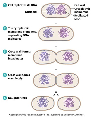
REPRODUCTION
It was earlier thought that the reproduction in bacteria is only asexual. But the recent studies revealed that the bateria also reproduce sexually.
ASEXUAL REPRODUCTION :
Asexual reproduction in Bacteria takes place by fission, budding and endospores.
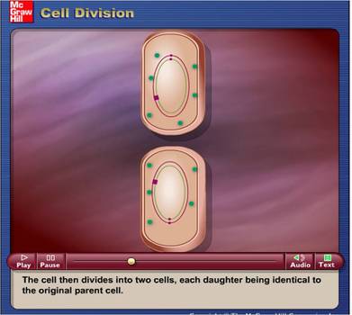
1. FISSION
- The fission is also called binary fission because in this process a bacterial mother cell splits into two daughter cells.
- In this process the circular DNA molecule first divides into two. In each of the two daughter DNA molecules one strand comes from the parent.
- The division of the nuclear material is followed by the cytoplasmic division.
- A peripheral ring of the plasma membrane invaginates and grows towards the centre to form a double transverse membrane.
- This newly formed wall in between the two halves thickens considerbly and splits into two, one for each daughter cell.
- The splitting takes place by the appearance of a constriction at the periphery and it slowly reaches the centre.
- Under favourable conditions fission occurs once in every 18-20 minutes.
|
Animation: Binary fission
|
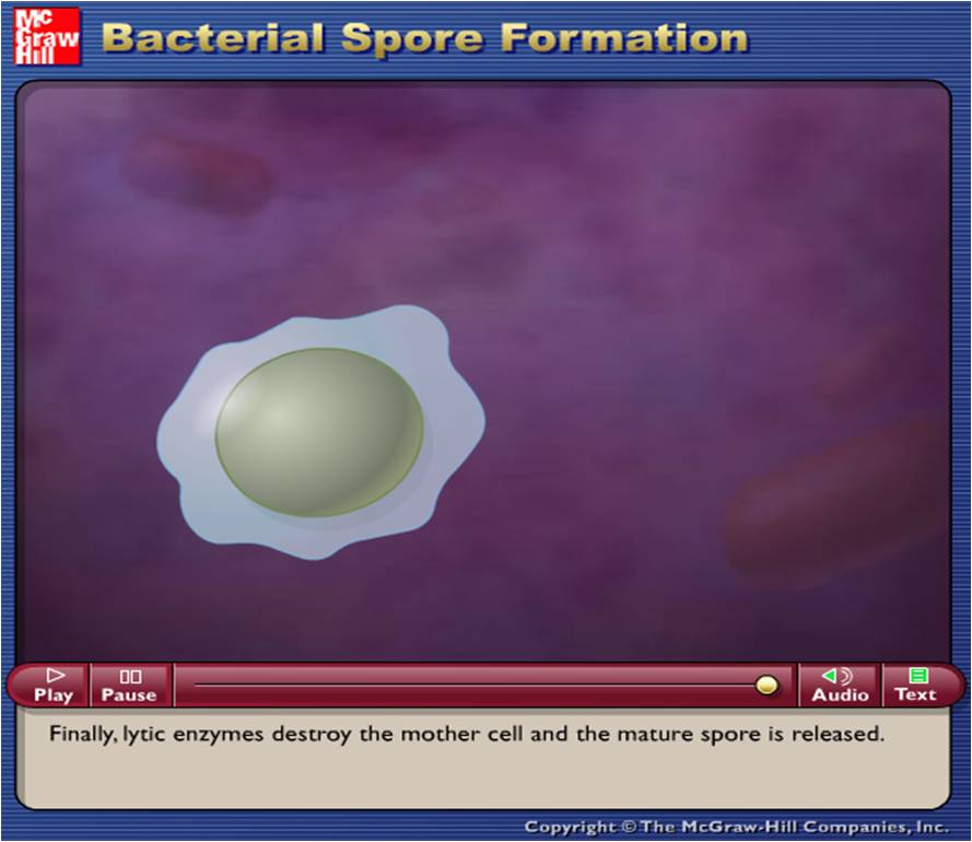
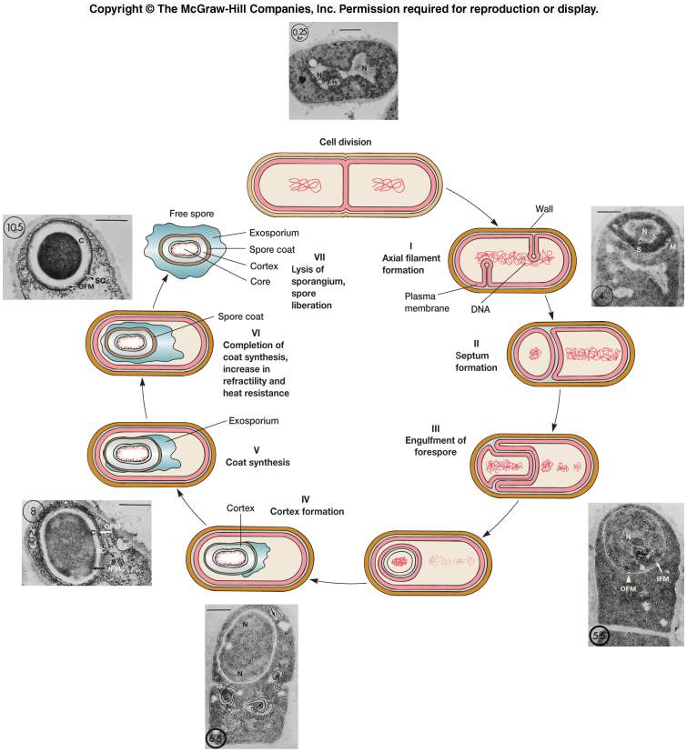
2. ENDOSPORE FORMATION (EXTRA INFORMATION)
|
Animation: Endospore formation
|
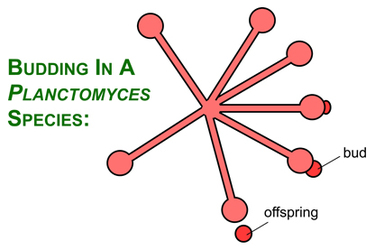
3. Budding
Budding has been observed in some members of the Planctomycetes, Cyanobacteria, Firmicutes and the prosthecate Proteobacteria. Although budding has been extensively studied in the eukaryotic yeast Saccharomyces cerevisiae, the molecular mechanisms of bud formation in bacteria are not known. A schematic representation of budding in a Planctomyces species is shown below.
Budding has been observed in some members of the Planctomycetes, Cyanobacteria, Firmicutes and the prosthecate Proteobacteria. Although budding has been extensively studied in the eukaryotic yeast Saccharomyces cerevisiae, the molecular mechanisms of bud formation in bacteria are not known. A schematic representation of budding in a Planctomyces species is shown below.
SEXUAL REPRODUCTION (Recombination) (EXTRA INFORMATION)
- Sexual reproduction is absent in bacteria because these do not form gametes.However, exchange of genetic material does take place which is known as recombination.
- Recombination-process by which one or more nucleic acid molecules are rearranged or combined to produce a new nucleotide sequence.
- In eucaryotes, recombination occurs during meiosis and results from crossing-over between homologous chromosomes (chromosomes containing identical sequences of genes)
- Types of recombination
- General recombination usually involves a reciprocal exchange in which a pair of homologous sequences breaks and rejoins in a crossover; nonreciprocal general recombination involves the incorporation of a single strand into the chromosome to form a stretch of heteroduplex DNA
- Site-specific recombination is the nonhomologous insertion of DNA into a chromosome; often occurs during viral genome integration into the host, a process catalyzed by enzymes specific for the virus and its host
- Replicative recombination accompanies replication and is used by some genetic elements that move about the genome
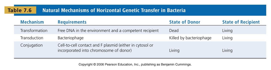
Methods of recombination
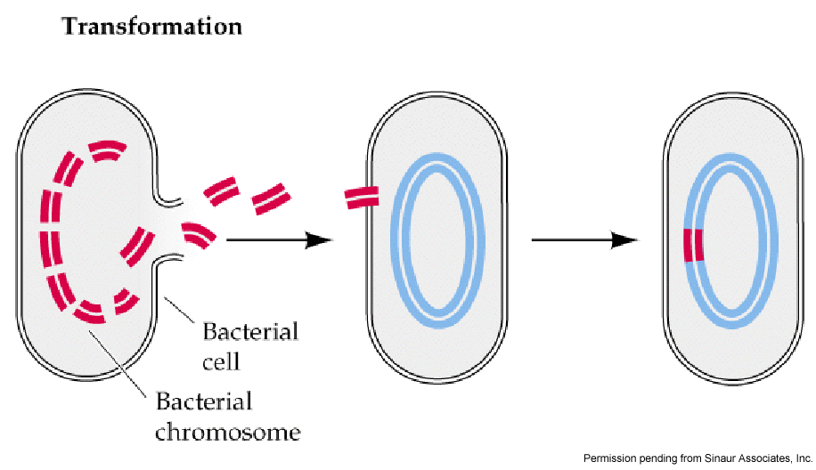
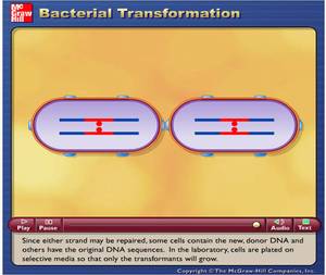
A. Transformation
Transformation is the process by which "naked" DNA is introduced into a bacterial cell. "Naked" DNA is generated when fragments of DNA or plasmids are released from damaged or dying cells. The fragments of DNA are then taken up by a living recipient cell and recombined with the recipient cell genome.
Transformation is the process by which "naked" DNA is introduced into a bacterial cell. "Naked" DNA is generated when fragments of DNA or plasmids are released from damaged or dying cells. The fragments of DNA are then taken up by a living recipient cell and recombined with the recipient cell genome.
|
ANIMATION
|
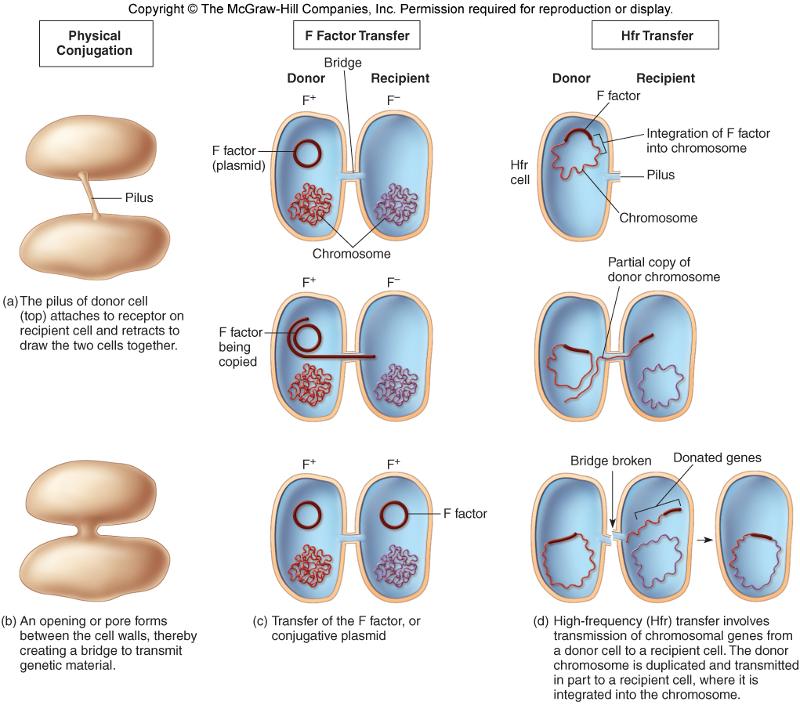
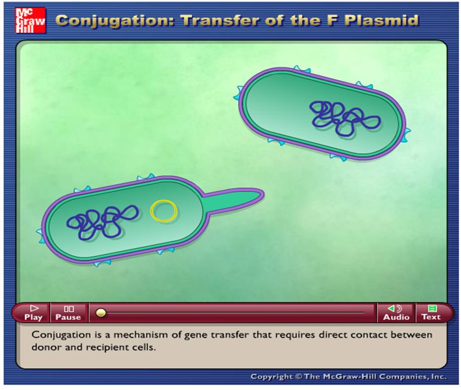
B. Conjugation
DNA from a donor cell is transferred to a recipient cell through a conjugation tube (pili).
DNA from a donor cell is transferred to a recipient cell through a conjugation tube (pili).
|
ANIMATION : CONJUGATION IN BACTERIA
|
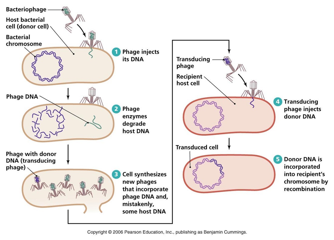
C. Transduction
Bacterial genes are carried from a donor cell to a recipient cell by a bacteriophage.
Bacterial genes are carried from a donor cell to a recipient cell by a bacteriophage.
|
ANIMATION : TRANSDUCTION

|
SUMMARY OF GENETIC RECOMBINATION
- Bacterial Plasmids
- Plasmids-small, circular DNA molecules that are not part of the bacteriumís chromosome
- Have their own replication origins, replicate autonomously, and are stably inherited
- Can be eliminated from a cell by a process called curing, which can occur either spontaneously or as a result of treatments that inhibit plasmid replication but do not affect host cell reproduction
- Episomes are plasmids that can exist either with or without being integrated into the host chromosome
- Conjugative plasmids usually have genes for sex pili and can transfer copies of themselves to other bacteria during conjugation
- Fertility factors-episomes that can direct the formation of sex pili and transfer copies of themselves during conjugation
- Resistance factors-(R plasmids); have genes for resistance to various antibiotics; some are conjugative; however, they usually do not integrate into the host chromosome
- Col plasmids-provide a competitive advantage to the bacteria having them; carry genes for the synthesis of bacteriocins (e.g., colicins) that are directed against other bacterial species; some col plasmids are conjugative and may also carry resistance genes
- Other types of plasmids include virulence plasmids, which make the bacterium more pathogenic by conferring resistance to host defense mechanisms or by carrying a gene for the production of a toxin, and metabolic plasmids, which carry genes for enzymes that utilize certain substances as nutrients (aromatic compounds, pesticides, etc.)
CYANOBACTERIA (BLUE GREEN ALGAE)
DISTRIBUTION
- Cyanobacteria are predominantly freshwater forms, a few are marine.
- The freshwater forms occur in ponds, lakes, pools and reservoirs. They impart unpleasant taste and smell to the water.
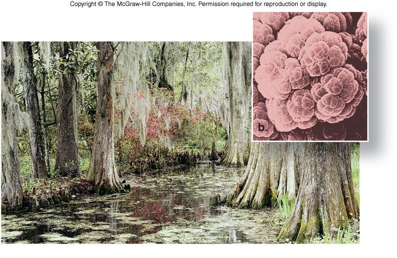
- One species of cyanobacteria containing red pigment (Trichodesmium erythraeum) flourishes in Red Sea and is responsible for the red colour of its water.
- Some grow in the soil and help in fixation of nitrogen.
- Some live in symbiotic relationship with other organisms.
FORMS
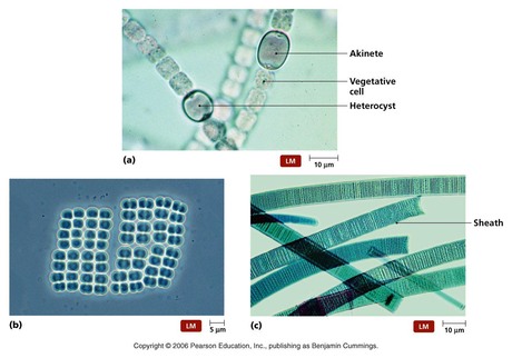
- The plant body is thallus-like.
- The thallus may be unicellular (as in Chrococcus, Anacystis ) colonial (as in Aphanocapsa, Gloeotricha) or filamentous (as in Oscillatoria, Nostoc, Scytonema, etc.).
- In Nostoc, some cells of the filament are differentiated to form heterocysts. Heterocysts are usually large-sized, rounded and thick-walled cells devoid of nucleoid. They are the sites nitrogen fixation.
- In Oscillatoria, filaments show oscillating motion and in Nostoc phormedium they show gliding movement.
|
CELL STRUCTURE
The cyanobacterial cell is normally larger than a bacterial cell. Like a bacterial cell, it consists of a tiny mass of protoplast surrounded by cell wall. It is differentiated into cell wall, cytoplasm and a nucleoid.
- Cell Wall: The cell wall completely surrounds the protoplast. It is a thin firm structure made up of peptidoglycan. External to the cell wall is a mucilaginous sheath. It has a great water absorbing and retaining capacity.
- Cytoplasm:
- The cytoplasm lacks ER, Golgi complex, mitochondria and lysosomes.
- The ribosomes are freely distributed in the cytoplasm and form polyribosomes during protein synthesis.
- The cytoplasm also contains photosynthetic pigments: the chlorophyll a and carotenoid. These occur in concentrically arranged flattened sacs, the lamellae called thylakoids. These forms have oxygen evolving photosynthetic system like higher plants, and produce oxygen (oxygenic photosynthesis).
- Cytoplasm also contains blue pigment phycocyanin and red pigment phycoervthrin. These two pigments are collectively known as phycobilin and are found inside small granules called cyanosomes or phycobilisomes.
- The reserve food is in the form of oil and fat droplets and proteinaceous granules.
- It lacks a definite nucleus. The nuclear material consists of a single chromosome made up of a naked strand of DNA helix which lies in the centre. DNA is not associated with histone proteins.
NUTRITION
- Because of the presence of chlorophyll a, cyanobacteria synthesise their own food from carbon dioxide and water in the presence of sunlight.This is called oxygenic photosynthesis.
- Certain cyanobacteria like Nostoc and Anabaena fix atmospheric nitrogen in the presence of oxygen. They are obligate photoautotrophs. They do not grow in darkness.
- Cyanobacteria are the earliest photosynthesisers which made the earth’s atmosphere aerobic. This provided the suitable condition for the evolution of aerobic bacteria and eukaryotes.
REPRODUCTION
- Cyanobacteria reproduce by vegetative and asexual means.
- Sex organs, gametes and flagellated zoospores are altogether absent.
- Vegetative reproduction takes place by fission and fragmentation.
- Unicellular forms multiply by binary fission.
- The filamentous forms reproduce by fragmentation into hormogonia (as in Nostoc and Oscillatoria). These serve as vegetative means of propagation.
- Some filamentous forms reproduce asexually by forming spores like akinetes, endospores, exospores.
- Endospores are formed endogenously in a vegetative cell as in Gloeocapsa.
- Exospores are produced exogenously as in Chamaesiphon.
- Akinetes are thick-walled spores with stored food as in Rivularia and Nostoc.
- SEXUAL REPRODUCTION :(PARASEXUALITY) Typical sexual reproduction is absent in cyanobacteria, but like bacteria combination by conjugation, transformation and transduction has been reported in some cases.
ECONOMIC IMPORTANCE OF CYANOBACTERIA
- Cyanobacteria are the earliest photosynthesisers. They developed oxygenic photosynthesis for the first time over 3 billion years back. These have changed the earth’s anaerobic atmosphere into aerobic atmosphere.
- Spirulina is cultivated in ponds and tanks as a protein-rich food for fish and other animals. It is also used as an important source of human food. Nostoc balls of terrestrial species are used as feed by Chinese and South Americans.
- Some cyanobacteria like Nostoc and Anabaena increase the soil fertility by fixing the free nitrogen of the atmosphere.These days, farmers inoculate rice fields with flakes of cyanobacteria to increase the yield.
- Cyanobacteria help in reclaiming barren soils and make them productive by increasing nitrogen, phosphorus, organic content and water-holding capacity of the soil. Examples: Anabaena, Aulosira, Scytonema, etc.
HARMFUL ACTIVITIES OF CYANOBACTERIA
- Some cyanobacteria produce toxins which are harmful to fishes and other aquatic animals. Such water is equally toxic to humans.
- Profuse growth of some cyanobacteria depletes oxygen content of waterbodies, thereby resulting in death of aquatic animals.
- Some species of cyanobacteria spoil the textiles, cordage and tents.
- Some cyanobacteria grow on the walls and roof of buildings during rainy seasons causing discolouration, leakage and corrosion of plaster and buildings.
- Lyngbya causes skin diseases.
- Some cyanobacteria, e.g., Oscillatoria, grow in municipal water reservoirs. They clog the water filters, and spoil the colour and taste by giving foul odour and fishy taste to water.
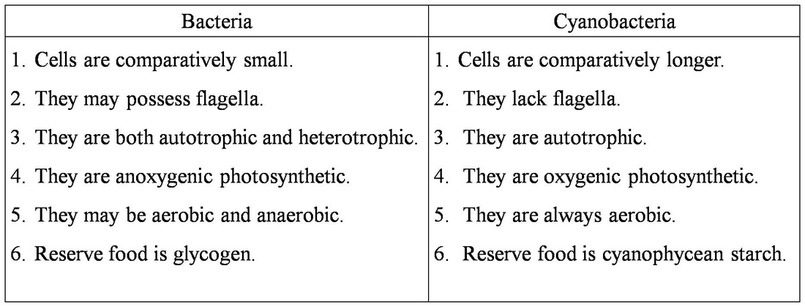
SIMILARITIES BETWEEN BACTERIA AND CYANOBACTERIA
- Both the groups lack a definite nucleus with nuclear membrane. Nuclear material is dispersed in the cytoplasm.
- Members of both the groups develop mucilage sheath around themselves.
- DNA and RNA not associated with nuclear proteins, the histones.
- Both the groups lack cell organelles like mitochondria, Golgi bodies, endoplasmic reticulum.
- Some members are able to tolerate extreme temperatures and desiccation.
- Some members carry fixation of nitrogen.
- Both the groups lack mitotic division.
- There is no sexual reproduction and meiotic division. There is complete absence of motile spores.
DIFFERENCES BETWEEN BACTERIA AND CYANOBACTERIA
MYCOPLASMA
INTRODUCTION
- Mycoplasmas are the simplest known aerobic prokaryotes without a cell wall.
- They are pleuropneumonia-like organisms (PPLO), discovered by Nocard and Roux (1898).
- Nowak (1929) placed them in genera Mycoplasma.
- They were first isolated from bovine sheep suffering from pleuropneumonia.
- Mycoplasma are considered to be intermediate between bacteria and viruses.
OCCURRENCE
- Mycoplasma occur in soil, sewage water, different types of substrates, humans, animals and plants.
- They are also found in hot water springs and other warm environments. They often contaminate tissue cultures rich in organic matter.
STRUCTURE
- Mycoplasmas show marked pleomorphism.
- Their size varies from 0.1—0.15 JIm.
- They lack a cell wall. Due to the absence of cell wall, they are plastic in nature and occur in various forms such as coccoid, granular, filamentous, cluster-like, etc.
- Because of variable shape, they are called ‘jokers of plant kingdom’.
|
LOCOMOTION, NUTRITION, REPRODUCTION
- Mycoplasmas are Gram negative and usually nonmotile. However, some forms show gliding movements.
- Mycoplasmas are heterotrophic. Some of them are saprophytes but most are parasitic on plants and animals.
- Mycoplasmas reproduce by budding and binary fission
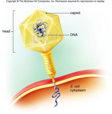
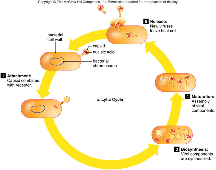
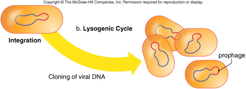
VIRUS
- Early Development of Virology
- Many epidemics of viral diseases occurred before anyone understood the nature of the causative agents of those diseases.
- Edward Jenner (1798) published case reports of successful attempts to prevent disease (smallpox) by vaccination; these attempts were made even though Jenner did not know that the etiological agent of the disease was a virus
- The word virus, which is Latin for poison, was used to describe diseases of unknown origin; filtering devices, which trapped bacteria but not viruses, were used by several scientists (Ivanowski, Beijerinck, Loeffler, Frosch, and Reed) to study a number of infectious agents; their recognition of an entity that was filterable (i.e., passed through a filter) led to the modern use of the term virus
- The role of viruses in causing malignancies was established by Ellerman and Bang (1908), who showed that leukemia in chickens was caused by a filterable virus, and Peyton Rous (1911), who showed that muscle tumors in chickens were caused by a filterable virus
- The existence of bacterial viruses was established by the work of Frederick Twort (1915), who first isolated bacterial viruses, and Felix díHerelle (1917), who devised a method for enumerating them and demonstrated that they could reproduce only in live bacteria
- W.M. Stanley (1935) helped demonstrate the chemical nature of viruses when he crystallized the tobacco mosaic virus and showed that it was mostly composed of protein; subsequently, F. C. Bawden and N. W. Pirie (1935) separated tobacco mosaic virus particles into protein and nucleic acid components