ICSE 10>BASIC BIOLOGY>1. CELL DIVISION
|
Scope of syllabus
Cell Division: Mitosis and its stages.
|
|
Helpful links
CHECK OUT SOME ANIMATIONS
http://www.cellsalive.com/mitosis.htm
http://www.maxanim.com/genetics/Mitosis/Mitosis.htm
http://www.sumanasinc.com/webcontent/animations/content/mitosis.html
QUIZ ON CELL DIVISION
http://www.syvum.com/cgi/online/oatm.cgi/squizzes/biology/mitosis.tdf?0
http://online.santarosa.edu/testbank/?11321
http://www.cellsalive.com/mitosis.htm
http://www.maxanim.com/genetics/Mitosis/Mitosis.htm
http://www.sumanasinc.com/webcontent/animations/content/mitosis.html
QUIZ ON CELL DIVISION
http://www.syvum.com/cgi/online/oatm.cgi/squizzes/biology/mitosis.tdf?0
http://online.santarosa.edu/testbank/?11321
What is cell division?
Cell division is a method by which new cells arises from pre-existing cells.
We can easily observe this when we get as small cut on our finger. The cut is healed and the scar will not be visible. This is the healing process.
Healing process is producing new cells.
The new cells produced should be exactly the same as their parent cell.
http://universe-review.ca/I10-04-cellnucleus.jpg
|
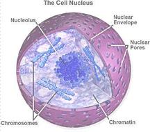
Chromosomes in the nucleus of the cell are the carriers of genetic information.
Every organism has a specific chromosome number. The number of these chromosomes should remain same inorder to maintain the cell structure and function. Thus when a cell divides, the daughter cells should receive the same chromosome number. Before beginning with the process of cell division let us get familiar with the chromosome structure. |
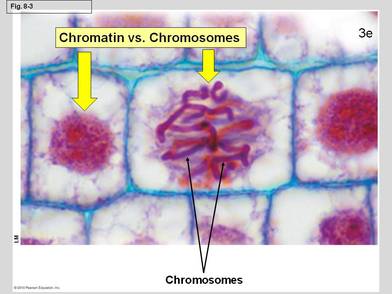
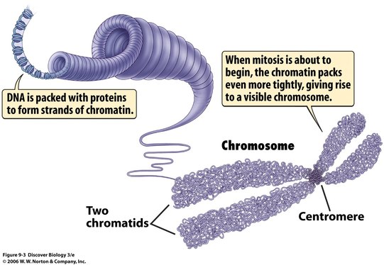
CHROMATIN AND CHROMOSOME
Nucleus contains thread like structures- chromatin
|
http://www.biology4kids.com/files/art/cell_nucleus2.gif
|
|
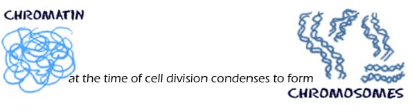
CHROMATIN
Chromatin is a mass of genetic material composed of DNA and proteins that condense to form chromosomes. |
CHROMOSOME
Chromosome is an organized structure of DNA and protein that is found in cells. |
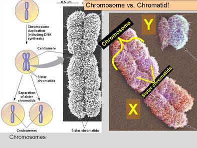
CHROMATID
Chromatid is the daughter strand of a duplicated chromosome that is joined by a single centromere. |
http://online.santarosa.edu/homepage/cgalt/BIO10-Stuff/Ch08-A-Mitosis/Chromatin-VS-Chromosomes.JPG
|
http://online.santarosa.edu/homepage/cgalt/BIO10-Stuff/Ch08-A-Mitosis/Chromosome-VS-Chromatid.JPG
|
|
Chromatid and DNAChromatid has a highly coiled of DNA and proteins. This thread like structure is called chromonema.
The DNA is coiled on proteins called histones. DNA coils around a core of eight histones forming a complex. This complex is called as nucleosome. |
Where is the DNA?
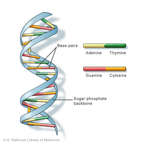
Structure of DNA
|
Discovery of DNA
James Watson and Francis Crick discovered the structure of DNA and were awarded the Nobel prize in 1962.
They constructed a double helix structure in 1953 DNA is the hereditary material present in humans and almost all organisms. (some viruses have RNA as genetic material)
Most DNA is present in the nucleus (nuclear DNA) and some DNA can be found in the mitochondria (mitochondrial DNA). |
http://evolution.berkeley.edu/evolibrary/images/history/dna_structure.gif
|
DNA is made up of two long parallel strands that are helically coiled around an axis.
The strands run in the opposite direction and is made up of repeating units called nucleotides. 
Structure of a nucleotide
Nucleotide has three sub units-
|
|
DOUBLE HELIX STRUCTURE-
|
DRAW THE DIAGRAM

|
DNA and chromosome
The DNA is packed with proteins to form chromosome.
Humans have 46 chromosomes. (23 pairs)
DID YOU KNOW
The record for minimum number of chromosomes belongs to a subspecies of the ant Myrmecia pilosula, in which females have a single pair of chromosomes.
The record for maximum number of chromosomes is found in found in the fern family. Ophioglossum reticulatum This fern has roughly 630 pairs of chromosomes or 1260 chromosomes per cell.
HOMOLOGOUS CHROMOSOME AND SISTER CHROMATID
http://www.phschool.com/science/biology_place/labbench/lab3/images/homologs.gif
|
HOMOLOGOUS CHROMOSOME-
These are chromosome pairs of approximately the same length, and centromere position. Out of the pair one is inherited from the mother (maternal) and one from the father (paternal). SISTER CHROMATID- These are two identical chromatids connected by a centromere. |
Chromosome Classification
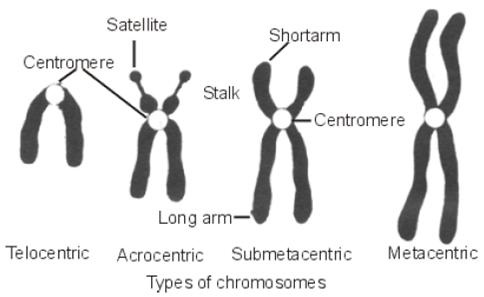
http://2.bp.blogspot.com/_fbXoeoCa5FQ/TGxfoW-eqxI/AAAAAAAAABk/OP8TaiYSEN0/s1600/16694_Types+of+chromosomes.JPG
|
Based on the position of the centromere, the chromosomes can be classified into four types
|
FUNCTION OF CHROMOSOME- carry genetic information from one generation to the other.
|
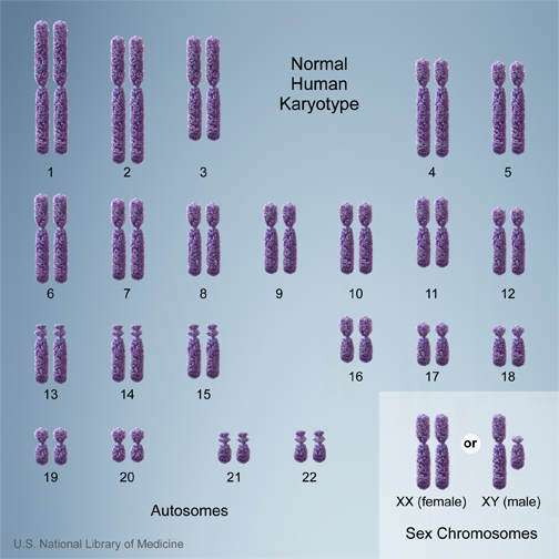
KARYOTYPE
When the chromosomes of a person are arranged in an organised manner it is called a Karyotype. Here the chromosomes are arranged and numbered by size, from largest to smallest. This arrangement helps scientists quickly identify chromosomal alterations that may result in a genetic disorder. |
|
Diploid and haploid
|
A cell having a single set of paired chromosomes is Diploid
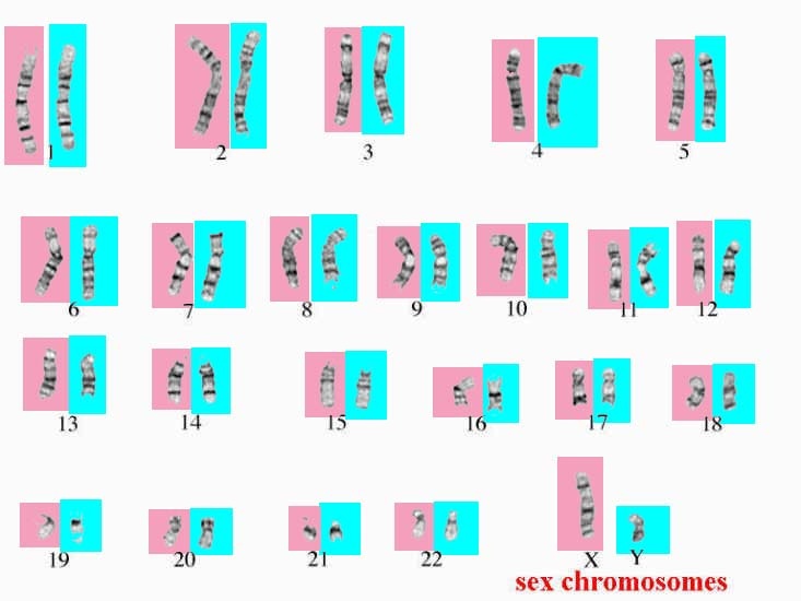
http://course1.winona.edu/sberg/IMAGES/human_karyotype2.jpg
|
A cell having a single set of unpaired chromosome is haploid
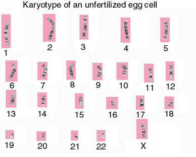
http://course1.winona.edu/sberg/IMAGES/human_karyotype3.jpg
|
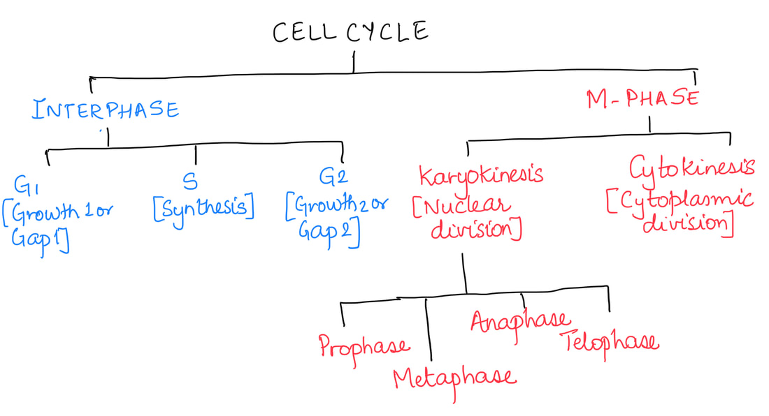
THE CELL CYCLE
|

http://virtuallaboratory.colorado.edu/BioFun-Support/AllGraphics/eukCycle.gif
|
G-1 post mitotic phase
|
S phase
|
G-2 pre mitotic phase
|
CELL DIVISION
|
Cell divides giving rise to two daughter cells that are identical to the parent cell and has the same chromosome number.
MITOSIS produces diploid cells |

http://www.daviddarling.info/images/cell_division.jpg
|
Cell divides giving rise to four daughter cells with half the number of chromosomes.
MEIOSIS produces haploid cells |
|
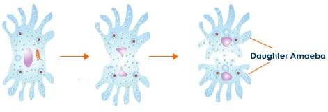
Sometimes there is direct cell division by simple cleavage of the nucleus, without spindle formation or the appearance of chromosomes.
This type of division is seen in prokaryotic cells and amoeba. This is called AMITOSIS. |
http://images.tutorvista.com/content/reproduction-in-animals/simple-binary-fission-amoeba.jpeg
|
Mitosis is also called- Equational division.
- Mitosis is an equational division in which a single cell divides to form two daughter cells.
- The daughter cells are identical to each other and the parent cell in all respect.
- Mitosis maintains the chromosome number i.e, the daughter cells have the same number of chromosome as the parent.
|
INTERPHASE
Growth period between two successive divisions.
|

|
|
PROPHASE
|

|
|
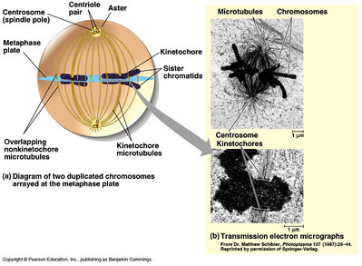
METAPHASE
|

|
|
The spindle apparatus, is a network of microtubules (also called "spindle fibers") that forms within a dividing eukaryotic cell, both during mitosis and meiosis
The kinetochore is the protein structure on chromatids where the spindle fibers attach during cell division to pull sister chromatids apart. Metaphase plate is a plane in the equatorial region of the spindle in dividing cells, along which the chromosomes become arranged during the metaphase. Kinetochore microtubules are spindle fibers that attach to the kinetochores and move the chromosomes to the center of the cell. Polar microtubules are not attached to chromosomes but overlap each other. Asters are short microtubules that radiate from the centrosomes. |
|
|
ANAPHASE
|

|
|
TELOPHASE
|

|
Drawing the stages of mitosis

|
|
Stages of mitosis as seen under microscope
In plant cell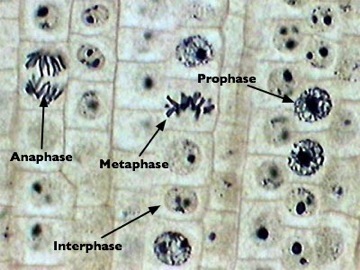
It occurs in the region of meristems.
Centrioles are not involved. Cytokinesis occurs by plate formation. cell plate or middle lamellae cements the daughter cells. |
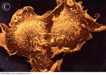
In animal cell
It occurs at several places.
Centrioles are involved in division. Cytokinesis occurs by cleavage. Cleavage creates intercellular space between daughter cells |
CYTOKINESIS
|
IN PLANT CELL
http://www.phschool.com/science/biology_place/biocoach/images/mitosisisg/mitcytpl.gif
|
IN ANIMAL CELL
http://images.tutorvista.com/content/feed/tvcs/cytokin.gif
|
|
What is the need for mitosis?
|
|
MEIOSIS is called the reductional division
|
|
FEATURES OF MEIOSIS-
Meiosis is a reductional division- chromosome number is reduced to half.
Pairing of homologous chromosomes- Synapsis
Chiasmata formation- Points at which non sister chromatids attach during synapsis
Crossing over- Exchange of genetic material between non sister chromatids.
Significance of meiosis-
Meiosis maintains definite and constant number of chromosomes in organisms.
Meiosis results in variation due to crossing over (exchange of genetic material)
Comparing mitosis and meiosis
|
|
|
|
|
sources of mitosis light micrographs-
http://www.anselm.edu/homepage/jpitocch/genbio/slidesother/inter92603.jpg
http://www.anselm.edu/homepage/jpitocch/genbio/slidesother/pro292603.jpg
http://jacusers.johnabbott.qc.ca/~biology/NYA/labs/NYALAB4/onionanaaphase.jpg
http://student.ccbcmd.edu/~gkaiser/biotutorials/dna/mitosis/images/metaphase_sa1_pc.jpg
http://schoolworkhelper.net/wp-content/uploads/2011/02/onion-mitosis-3-lab.jpg
http://www.anselm.edu/homepage/jpitocch/genbio/slidesother/telo292603.jpg
http://www.phschool.com/science/biology_place/labbench/lab3/images/anifield.gif
