ISC 12> STRUCTURE AND FUNCTION OF ANIMALS> 10. EMBRYONIC DEVELOPMENT
SCOPE OF SYLLABUS
physico-chemical events during fertilisation,
implantation, embryonic development up to blastocyst formation,
important features of human embryonic development (formation of heart, limbs, digits, appearance of hair on head, eyelashes, separation of eye lids, external genital organs and first movement of foetus with reference to time period)
placenta and its functions.
Parturition; lactation – hormonal control and importance.
Phases of embryonic development
|
|
A study of the development of an organism from fertilisation to the formation of young one is known as embryogeny.
When the male and female gamete fuse at the time of fertilisation, the resulting cell- the zygote has the full or diploid number of chromosomes. The structural changes in a zygote leading to the formation of adult form is embryonic development. During this a single celled egg transforms into a multicellular and fully formed organism. The phases include- 1. Gametogenesis 2. Fertilisation 3. Early development- including- cleavage, blastulation and gastrulation. 4. Differentiation and organogenesis. 5. Differentiation and morphogenesis. |
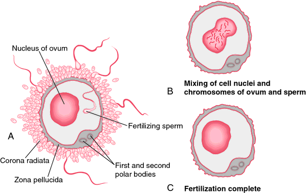
Fertilisation- stages
The fusion of haploid male gametes and female gametes to form zygote is called fertilisation. In humans the fertilisation is internal and takes place in the proximal part of the fallopian tube. The process of fertilisation involves different stages-
|
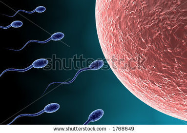
1.Approach of sperm to ovum
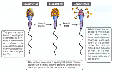
A single ejaculate of sperms contains around 200-400 million sperms in approximately 3 to 3.5 ml of semen. However only sperms are able to reach the fallopian tube. This is because of- acidity of the female genital tract, some are engulfed by leucocytes of vaginal epithelium, some are trapped in the cilia of the fallopian tube. 2. Capacitation The changes in the mammalian sperm that make it to fertilise the ovum are called capacitation. The main changes taking place are-
|
|
|
3. Agglutination reaction- The ovum secretes a chemical substance called fertilizin. It is a glycoprotein that has a number of spermophilic sites on its surface for the antifertilizin present on the head of sperm.
The adhesion of spermatozoa to the surface of egg is brought about by linking of fertilizin and anti fertilizin molecules. This reaction causes agglutination of sperm and thins out the number of sperm reaching the egg to reduce the chances of polyspermy. The adhesion of sperm to the egg of the same species through chemical recognition is known as agglutination. The process of fertilisation involves-
|
|
|
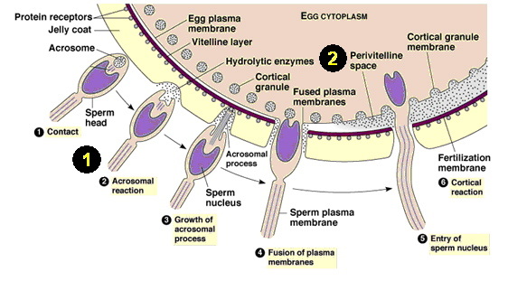
4. Penetration of sperm into the ovum and acrosome reaction
The ovum at the secondary oocyte stage is extruded from the ovary along with corona radiata cells. These follicular cells are glued together by a mucopolysaccharide, the hyaluronic acid. The sperm produces enzymes that are known as sperm lysins. The hyaluronidase enzyme helps to dissolve hyaluronic acid polymers in the intercellular spaces which hold granulosa cells of corona radiata together; corona penetrating enzyme dissolves the corona radiata and acrosin which dissolves the zona pellucida. The nucleus and the cytoplasm of the sperm are drawn in the ovum. Due to this acrosomal reaction the plasma membrane of the sperm fuses with the plasma membrane of the secondary oocyte so that the contents of the sperm enter the oocyte. This induces depolarisation of the oocyte plasma membrane and thus ensures monospermy. |
|
5. Cortical reaction
Due to this acrosomal reaction the plasma membrane of the sperm fuses with the plasma membrane of the secondary oocyte so that the contents of the sperm enter the oocyte. After fusing of sperm with egg membrane, cortical granules release chemicals into the perivitelline space that harden the zona pellucida. This prevents polyspermy.
Due to this acrosomal reaction the plasma membrane of the sperm fuses with the plasma membrane of the secondary oocyte so that the contents of the sperm enter the oocyte. After fusing of sperm with egg membrane, cortical granules release chemicals into the perivitelline space that harden the zona pellucida. This prevents polyspermy.
|
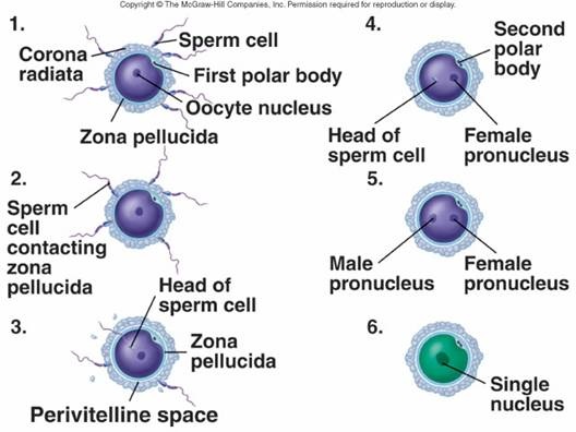
6. Activation of ovum
The entry of sperm induces a chemical signal in the egg cell. This initiates the formation of a fertilisation membrane around the egg. The cortical granules present beneath the plasma membrane of the oocyte fuse with the membrane and release their contents between the plasma membrane and the zona pellucida. This hardens the zona pellucida and further prevents the entry of other sperms in the egg cell. The secondary oocyte completes the second meiotic division producing ovum and second polar body. The second polar body degenerates immediately. 7. Fusion of sperm and egg nucleus.
The point of contact of the sperm entering the oocyte is called as the cone of reception or the fertilisation cone. The sperm nucleus now moves towards the egg nucleus along a definite path called copulation path. This leads to the fusion of the male and female pronucleus called fertilization. The fertilized egg is now called zygote. |
|
|
SIGNIFICANCE OF FERTILIZATION:
|
|
Post fertilization changes
|
The entrance of sperm nucleus along with its centriole in the egg cytoplasm induces formation of spindle and prepares the zygote for the first nuclear division.
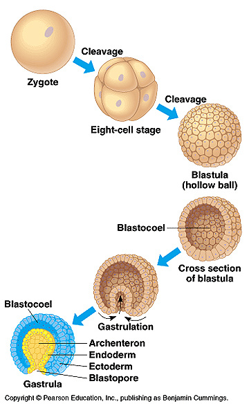
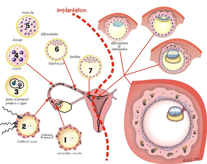
The division occurring in the zygote is called cleavage. It is an embryological process that transforms a single fertilized egg cell (zygote) into a hollow, spherical and a multi-cellular stage - blastula. The mammalian zygote undergoes holoblastic and equal cleavage. The cleavage occurs in the fallopian tube as the zygote passes towards the uterus. 30 hrs after fertilisation: The first cleavage occurs. This results in the formation of two blastomeres, one slightly bigger than the other. 60 hrs after fertilisation: The second cleavage occurs. The larger blastomere divides a little sooner than the smaller one thus there is a transitory three celled stage before the formation of the four celled stage. 72 hrs after fertilisation: The third cleavage takes place. Subsequent cleavage divisions follow to form a solid ball of cells. The cleavage leads to increase in cell number, without growth of cells. The blastomeres formed become smaller and smaller. Morula- The stage that consists of 16-32 cells and is of the same size as the zygote. Morula reaches the uterus about 4-6 days after fertilisation. The morula consists of centrally located cells, the inner cell mass and a surrounding layer, the outer cell mass. The inner cell mass gives rise to the embryo proper whereas the outer cell mass forms the trophoblast, that later develops to form the placenta. |
|
Blastulation
The rearrangement of the blastomeres result in the differentiation of morula to blastocyst.
As these cells continue to divide, a cavity appears inside the morula. This sphere of cells with a cavity in the center is called as blastula or blastocyst. The cavity developed inside is called as the blastocoel.
As the blastocyst formation takes place, the zona pellucida starts to disintegrate, this facilitates the implantation and rapid growth of the embryo.
The process by which the blastocyst comes out of the zona pellucida is called hatching. The presence of zona pellucida prevents implantation at unfavourable sites.
The layer of cells which form the outer wall of the blastocyst is called trophoblast or trophoectoderm. The blastomeres on this layer form the protective and trophic membranes. They develop to form the foetal part of the placenta and provide nutrition to the developing embryo.
The inner cell mass gives rise to the embryo proper. The part of the blastocyst to which the inner cell mass is attached is the animal pole or embryonic pole.

|
|
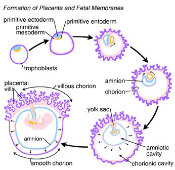
Implantation
The attachment of the blastocyst to the uterine wall is called implantation. This takes place about 5 to 7 days after ovulation. The direct contact of the blastocyst with the uterine endometrium stimulates the trophoblast cells at the embryonal pole to divide rapidly. These cells release proteolytic enzymes that erode the cells of endometrium. The blastocyst now sinks into the pit formed in the endometrium. The endometrium then grows around the blastocyst.
The trophoblast forms the villi to obtain nourishment. The trophoblast invading the blastocyst develops to form the chorion an extra-embryonic membranes.
The trophoblast forms the villi to obtain nourishment. The trophoblast invading the blastocyst develops to form the chorion an extra-embryonic membranes.
|
|
|
The chorionic cells and human chorionic gonadotropin
The chorionic cells of the placenta secrete a hormone - human chorionic gonadotropin (HCG). It carries out the following function-
- Maintains corpus luteum.
- Stimulates secretion of progesterone by the corpus luteum.
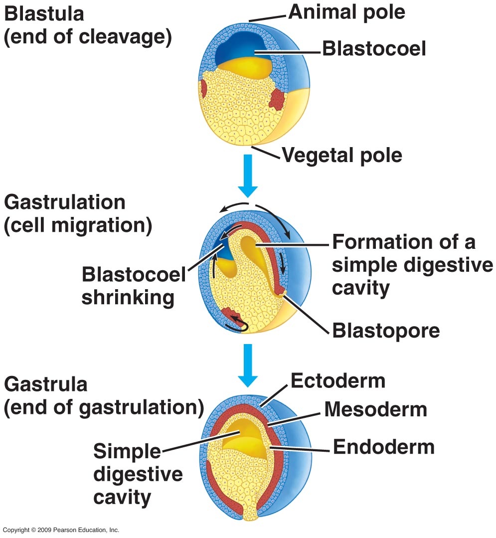
Gastrulation : formation of three germinal layers
|
The undifferentiated blastomeres move in small masses to form sheets of cells. These cells differentiate into three germ layers- ectoderm, mesoderm and endoderm.
These are called primary germ layers. These movements are called morphogenetic movements. The over all event is called gastrulation. Gastrulation can be defined as a dynamic process in the development that involves the movement of prospective organ forming areas from the blastodermic vesicle to their definitive positions in the embryo and their reorganisation into three primary layers. These primary germ layers give rise to-
|
|
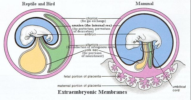
EXTRA-EMBRYONIC MEMBRANES
|
There are thin layers of tissue that surround the growing embryo. These are extra embryonic membranes that provide protection and nourishment to the developing foetus.
These are-
The amnion and the amniotic cavity enlarge and surround the embryo.
The lining of allantois fuses with the somatic mesoderm of the chorion to form the allanto-chorion. This layer then develops to form the placenta.
The role of amniotic fluidThe amniotic fluid-
1. Cushions the developing embryo- provides protection against shocks and jerks. 2. Acts as a lubricant- allows movement of the limbs. 3. Prevents desiccation of the embryo. 4. Maintains a consistent temperature. |
|
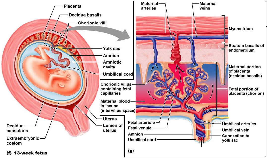
PLACENTA
It is a temporary organ formed in the placental mammals that is derived from two different individuals. It is an intimate connection between the foetus and the mother.
There are large finger like projections called villi. The uterine wall that in contact with the villi forms depressions called crypts.
The part of the placenta contributed by the foetal membrane of the foetus is called foetal placenta and the part contributed by the mother's uterine wall is the maternal placenta.
In the placenta the foetal blood comes in close contact with maternal blood that facilitates the exchange of materials.
Functions of placenta-
- transport of nutrients from the maternal blood to the foetus.
- helps in respiratory exchange
- removal of nitrogenous wastes from the foetus into the maternal blood stream.
- antibodies from the mother passes to the foetus and provides passive immunity.
- It helps in storage of fat, glycogen etc.
- produces hormones like progesterone, estrogen, gonadotropins etc.
- certain pathogens and drugs also get transported from the maternal blood stream to the foetus, that can lead to problems.
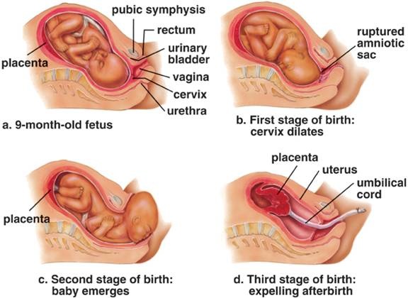
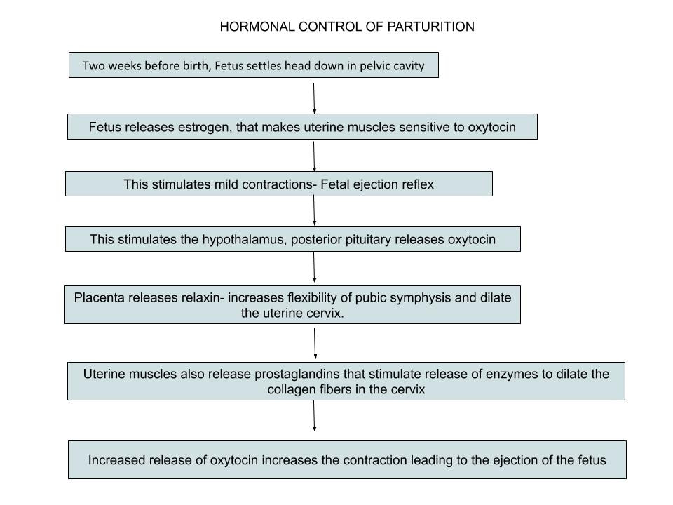
PARTURITION OR BIRTH
The act of expelling the foetus after a complete term of pregnancy or gestation.
The gestation period is completed in 280 days.
Parturition involves forceful muscular contractions called labor.
The head of the foetus settles down into the pelvic cavity two weeks before birth. When gestation is completed, labor starts and the foetus is expelled out.
The onset of labor commences with the rupture of the amniotic sac and leakage of the amniotic fluid.
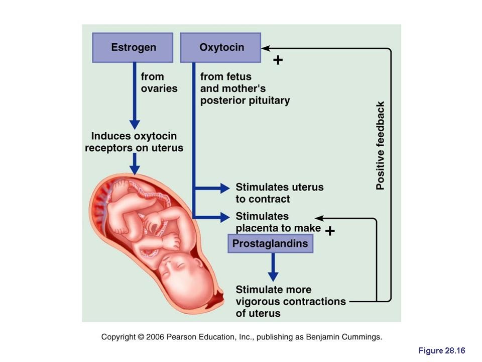
Role of hormones in parturition
The hormone Oxytocin causes contractions during parturition.
The hormone Relaxin secreted by the placenta relaxes the cervix and pelvic ligaments for easy birth. (Relaxin is also secreted by the corpus luteum in non pregnant females)
The hormone Relaxin secreted by the placenta relaxes the cervix and pelvic ligaments for easy birth. (Relaxin is also secreted by the corpus luteum in non pregnant females)
|
|
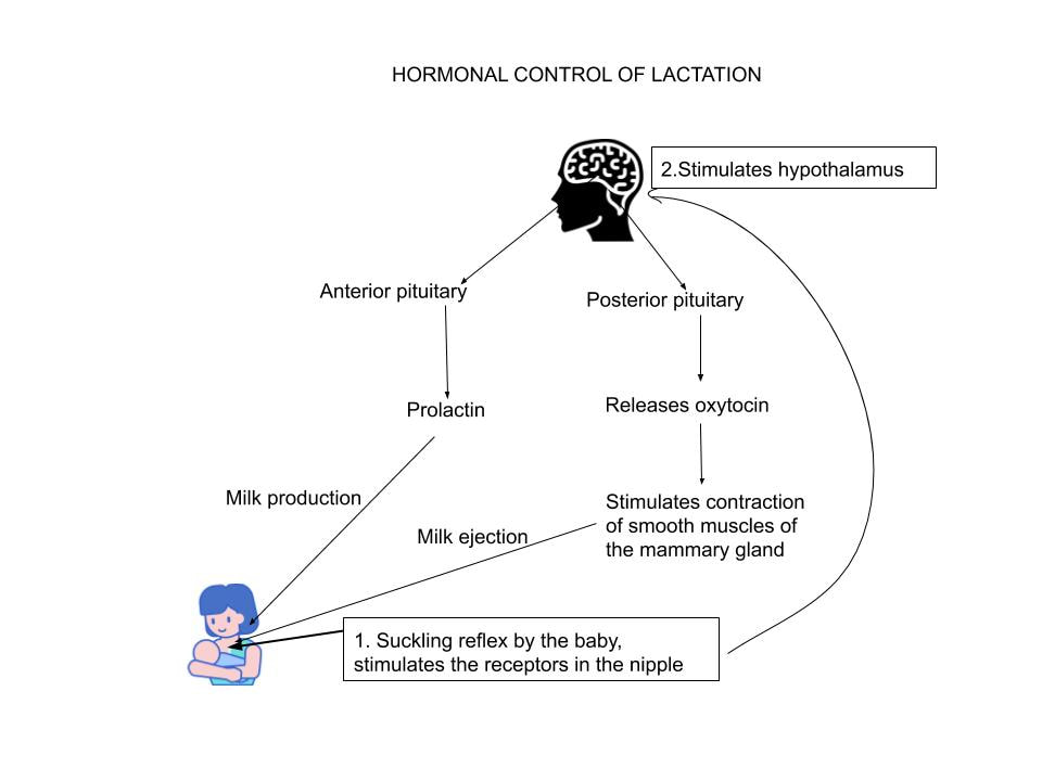
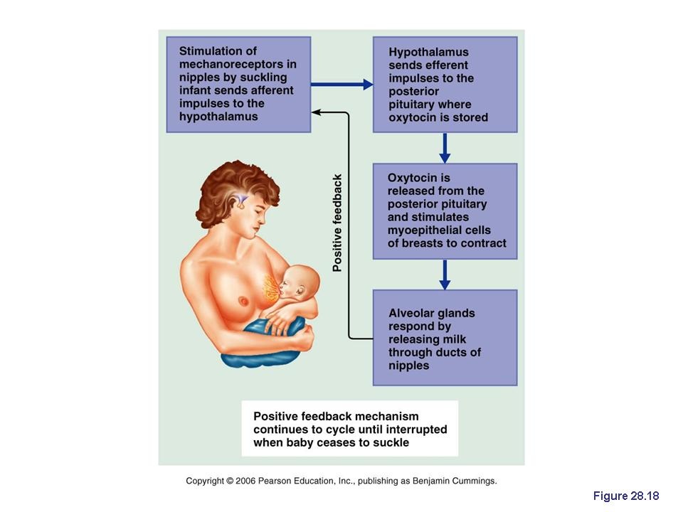
Lactation
|
The secretion of milk from the mammary gland and the period of time the mother lactates is called lactation.
During pregnancy the size of mammary glands increases. After birth the hormone prolactin stimulates the mammary glands to produce milk. The stored milk is released after parturition. The suckling of the nipples by the baby causes the release of the milk. Milk secretion begins usually within 24 hrs under the influence of prolactin. The first fluid that is released after birth is called colostrum. Colostrum is special milk that is yellow in color and thick and sticky. It is low in fat, and high in carbohydrates, protein, and antibodies. It is extremely easy to digest. Colostrum has a laxative effect on the baby, helping him pass his early stools, which aids in the excretion of excess bilirubin and helps prevent jaundice. |
|
Role of hormones in lactation
|
|
Multiple births
Normally a human female gives birth to one child at a time. However at times two or more babies are born.
Such babies are called twins or multiple births.
Twins can develop in two ways-
Monozygotic or identical twins- arise from single egg.
Dizygotic or fraternal twins- two eggs fertilised by two different sperms.
Such babies are called twins or multiple births.
Twins can develop in two ways-
Monozygotic or identical twins- arise from single egg.
Dizygotic or fraternal twins- two eggs fertilised by two different sperms.
