ISC 11>CONTENT>U-2. DIVERSITY OF LIFE> 8.MORPHOLOGY AND ANATOMY OF COCKROACH
SCOPE OF SYLLABUS: Morphology and anatomy of different systems of cockroach.
Elementary knowledge of the digestive, respiratory, circulatory, excretory, nervous and reproductive system.
Elementary knowledge of the digestive, respiratory, circulatory, excretory, nervous and reproductive system.
PRE-LEARNING VIDEO
http://www.youtube.com/watch?v=daYvNM2IuOg
CLICK TO WATCH ON YOUTUBE
CLICK TO WATCH ON YOUTUBE
DISSECTING THE COCKROACH- PRACTICAL |
CLASSROOM PRESENTATION |
CLASSIFICATION
Blatta orientalis, B germanica found in India |
|
MORPHOLOGY OF COCKROACH
Dorsal view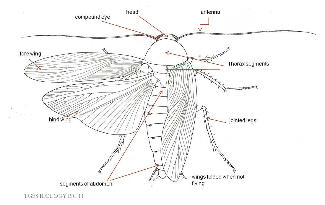
|
Ventral view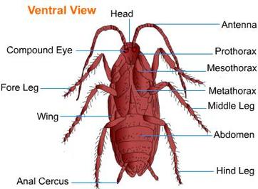
|
External features
|
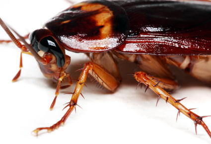
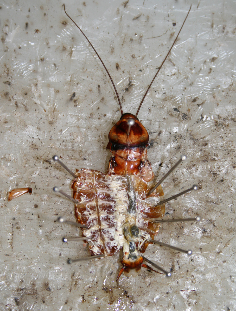
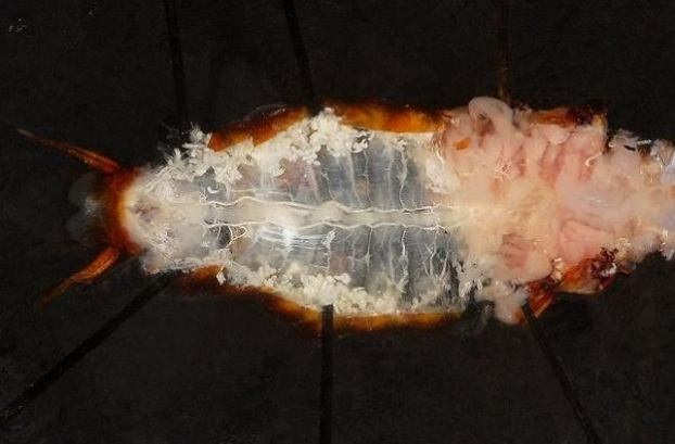
The body of the cockroach is elongated and segmented.
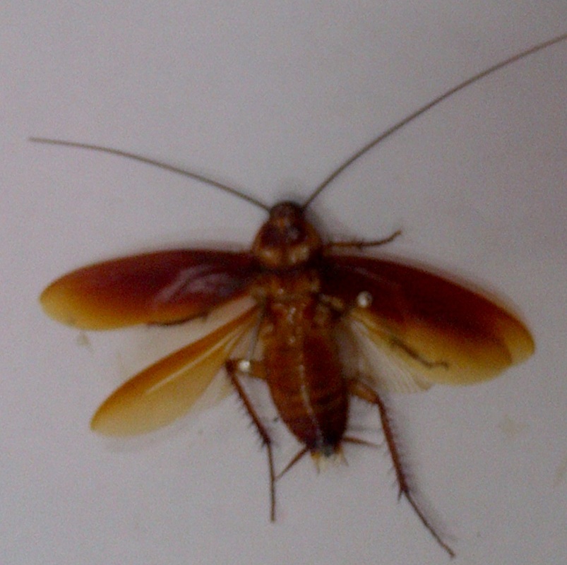
It is dark brown or reddish brown in colour. The exoskeleton is thick and hard made up of calcareous plates called sclerites. There are 10 segments. The segments on — on dorsal side (or notum) are called Tergum —on ventral side are called Sternum. The exoskeleton is coated with wax impermeable to water. It protects the body from loss of water and provides rigidity and surface for attachment of body muscles. The adjacent segments are joined by thin, soft and flexible arthroidal membrane. The body is divisible into head, thorax and abdomen. The cockroach has three pairs of jointed appendages and two pairs of wings. The fore wings are mesothoracic and are called wing covers or tegmina or elytra. They cover the hindwings and are protective in function. These are dark stiff opaque and leathery. The hind wings are large, thin, membranous and transparent. They are kept folded below the tegmina and are used for flying. |
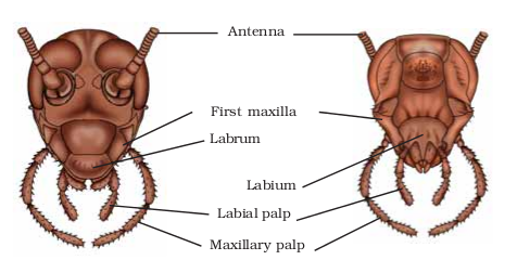
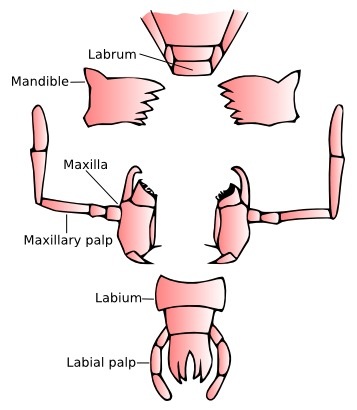
Mouth parts of cockroach
|
Ventrally, an opening called mouth is present on the head that remains surrounded by the mouth parts consisting of a pair of mandibles, first maxillae, labium or fused second maxillae, hypopharynx and labrum.
The mouth parts of the cockroach help in 'biting and chewing' its food. Functions of the mouth parts:
|
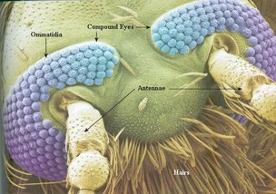
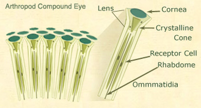
COMPOUND EYE
|
|
The compound eyes are situated at the dorsal surface of the head. Each eye consists of about 2000 hexagonal ommatidia (sing.: ommatidium). With the help of several ommatidia, a cockroach can receive several images of an object. This kind of vision is known as mosaic vision with more sensitivity but less resolution, being common during night (hence called nocturnal vision).
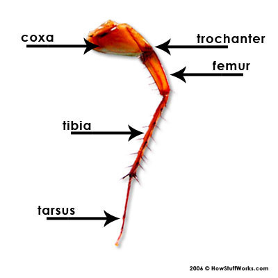
Leg of cockroach
|
A cockroach's thorax attaches three pairs of legs. Each of the three pairs of legs is named after the region of the thorax to which it attaches:
The prothoracic legs are closest to the cockroach's head. These are the shortest legs, and they act like brakes when the roach runs. The middle legs are the mesothoracic legs. They move back and forth to either speed the roach up or slow it down. The very long metathoracic legs are the cockroach's back legs, and they move the cockroach forward. These three pairs of legs, are substantially different in lengths and functions, but they have the same parts and move the same way. The upper portion of the leg, called the coxa, attaches the leg to the thorax. The other parts of the leg approximate parts of a human leg: The trochanter acts like a knee and lets the cockroach bend its leg. The femur and tibia resemble thigh and shin bones. The segmented tarsus acts like an ankle and foot. The hook-like tarsus also helps cockroaches climb walls and walk upside down on ceilings. |
|
POSTERIOR ABDOMINAL SEGMENTS
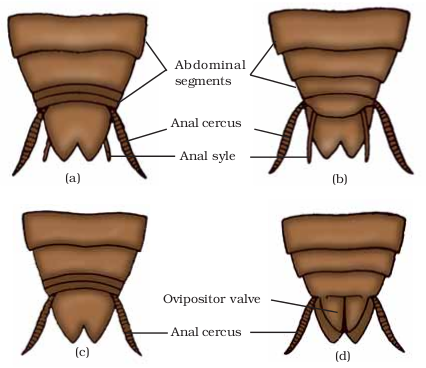
The figure shows posterior abdominal segments of cockroach (a) Male dorsal view (b) Male ventral view (c) Female dorsal view (d) Female ventral view.
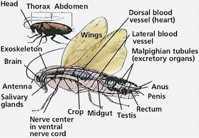
Side view of cockroach showing the location of the systems
DIGESTIVE SYSTEM
|
|
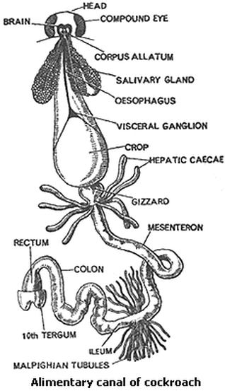
- The alimentary canal is long and somewhat coiled divisible into three main parts namely foregut, midgut and hindgut.
- Foregut (stomadaeum) is differentiated into five parts: Buccal chamber, pharynx, oesophagus, crop and gizzard.
- Gizzard is muscular and internally provided with six cutical teeth which crushes the food.
- A stomodaeal valve is present between gizzard and mesenteron.
- Midgut (mesenteron or ventriculus) is short, tubular lined with glandular endoderm.
- At anterior end of mesenteron there are eight blind glandular hepatic caecae which secrete digestive enzymes.
- Hindgut (proctodaeum) comprises ileum, colon and rectum.
- The wall of rectum is provided with six rectal papillae. They help in the absorption of water and salts.
- Cockroach is omnivorous feeds on all sorts of organic debris.
- The digestive enzymes of saliva are mainly zymase and amylase.
- Most of the nutrients of food are digested in the crop.
- Absorption of digested food takes place in mesenteron.
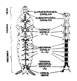
NERVOUS SYSTEM
|
|
It consists of:
- Central Nervous System
- Peripheral Nervous System
- Sympathetic or Visceral System
- Central Nervous System: It consists of brain or supra oesophageal ganglion. Brain gives off a pair of short, stout cords, the circumoesophageal connectives, that encircle the oesophagus and pass downwards and backwards over the suboephageal ganglion situated below oesophagus.
From the sub esophageal ganglion passes backwards into the thorax, a double ventral nerve-cord, which bears three ganglia in the thorax and six in the abdomen. - Peripheral Nervous System: It consists of nerves, which are given off from the ganglia so as to innervate all the parts of the body.
- Sympathetic or Stomatogastric Visceral Nervous System: It consists of a frontal ganglion, which is situated on the dorasl side of the oesophagus in the head. From this ganglion, a median unpaired recurrent nerve reaches the visceral ganglion situated on the crop. Various nerve branches are given off from the visceral ganglion.
The frontal ganglion is jointed with the central nervous system by nerves, which connect the circumoesophageal commissures.
Sensory structures
- Thigmoreceptors: They are receptors of touch. Thigmoreceptors are present on body, antenna, maxillary palps and legs.
- Olfactory receptors: They receive various smells. Olfactory receptors are present on antenna and palps.
- Gustatory receptors: They are for sense of taste. Gustatory receptors are present on maxilla and labial palps.
- Thermoreceptors- detect changes in temperature, present on the pads between the first four tarsals.
- Auditory receptors- for hearing, present on the anal cerci respond to air or earth borne vibrations.
CIRCULATORY SYSTEM
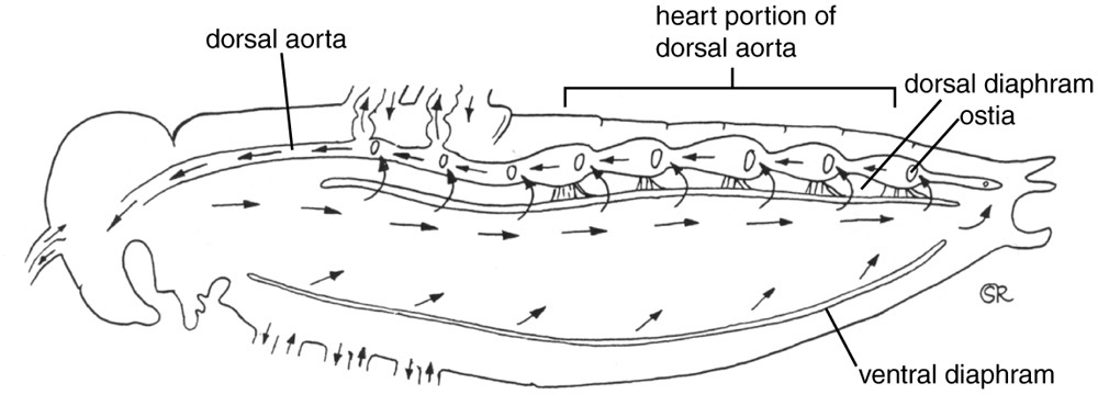
|
Blood vascular system is open and lacunar type. Body cavity contains blood, which bathes viscera in it therefore known as Haemocoel.
Blood vascular system consists of a tubular heart, a blood vessel called anterior aorta and a system of ill defined blood spaces or sinuses. |
The Blood Sinuses
The large body cavity or haemocoel is divided by two membranous horizontal partitions, into three wide and flattened sinuses-the dorsal pericardial sinus containing the 'heart', the middle perivisceral sinus containing the gut, and the ventral perineural sinus or sternal sinus containing the nerve cord. The partition between pericardial and perivisceral sinuses is called dorsal diaphragm and between perivisceral and perineural sinuses is called ventral diaphragm. The sinuses intercommunicate by pores in the respective diaphrams. A pair of fan like, triangular alary muscles in the floor of the pericardial sinus in each segment reinforce the dorsal diaphrams by their broad bases and also connect it, by their pointed tips with the tergite of the segment.
Circulation of Haemolymph
The pumping force that propels the haemolymph is provided by the pulsations of the 'heart'. The respiratory movements of abdomen and contraction of alary muscles increase this force.
- From the pericardial sinus, the haemolymph enters into heart through ostia.
- The valve like ostia close, preventing back flow of haemolymph into the pericardial sinus. Therefore, some of its haemolymph is pumped into segmental vessels, while most of its poured into the head sinus through the terminally opening anterior aorta.
- From the head sinus, the haemolymph flows backward into the thorax and abdomen. While flowing backwards from head sinus, the haemolymph remains in the ventral part due to presence of Oesophagus in dorsal part and so it fills into the perineural sinus.
- From the perineural sinus, the haemolymph, now, flows into the perivisceral sinus through the pores of ventral diaphram in abdominal region.
- Then from perivisceral sinus, it flows into pericardial sinus through the pores of dorsal diaphram. Then, during heart's diastole, it fills in the heart through the ostia.
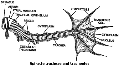
Respiratory System
|
The respiratory system consists of a network of trachea, that open through 10 pairs of small holes called spiracles present on the lateral side of the body. Thin branching tubes (tracheal tubes subdivided into tracheoles) carry oxygen from the air to all the parts. The opening of the spiracles is regulated by the sphincters. Exchange of gases take place at the tracheoles by diffusion.
|
|
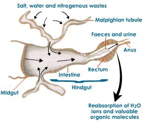
Excretory System
|
Excretion is performed by Malpighian tubules.
Each tubule is lined by glandular and ciliated cells. They absorb nitrogenous waste products and convert them into uric acid which is excreted out through the hindgut. Therefore, this insect is called uricotelic. In addition, the fat body, nephrocytes and urecose glands also help in excretion. |
|
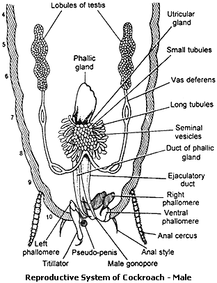
Reproductive System of Cockroach - Male
|
In cockroach, sexes are separate, so dioecious.
|
|
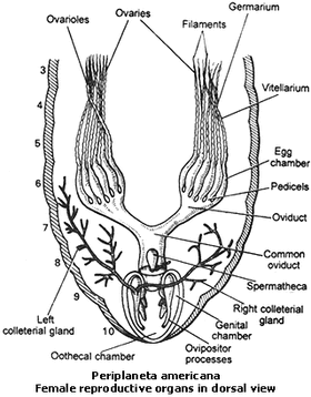
Reproductive System of Cockroach - Female
|
|
- Nymph of cockroach emerge out from ootheca. A nymph resembles to adult in general structure but lacks the wings and mature reproductive organs.
- Instar a large stage in the development of insects (larval instar, nymphal instar). Period between two moults in insects is termed stadium.
- In periplaneta americana the nymph grows by moulting about 13 times to reach the adult from blatta orientalis moults 6 times.
- Metamorphosis is regulated by two hormones, ecdysone secreted by prothoracic glands and juvenile hormone secreted by corpora allata.
