BIOLOGY4ISC
  • HOME
  • ICSE 10
    • SYLLABUS
    • CONTENT >
      • Basic Biology >
        • 1. Cell division
        • 2. Genetics
      • Plant physiology >
        • 3. Osmosis and Ascent of sap
        • 4. Transpiration
        • 5. Photosynthesis
      • Human anatomy and physiology >
        • 6. Circulatory system
        • 7. Excretory system
        • 8. Nervous system and sense organs
        • 9. Endocrine system
        • 10. Reproductive system
      • Physical health and hygiene >
        • 11. Human population
        • 12. Aids to health and health organisation
    • PAPERS
  • ISC 11
    • SYLLABUS
    • CONTENT >
      • U-1 Diversity of life >
        • a. Biological classification >
          • Kingdom Monera
          • Kingdom Protista
          • Kingdom Fungi
        • c. Kingdom Plantae >
          • Thallophyta
          • Bryophyta
          • Pteridophyta
          • Gymnosperms
          • Angiosperms
        • d. Kingdom Animalia
      • U-2:Structural organisation in plants and animals >
        • b. Anatomy of flowering plants >
          • Basic idea of how secondary growth
          • Plant tissues
        • c. Structural organisation in animals >
          • Animal tissues
          • Connective tissue
          • Morphology and anatomy of cockroach
      • U-3. Cell structure and function >
        • a. Cell : unit of function >
          • cell organelles
          • nucleus
          • Cell membranes
        • b. Biomolecules >
          • Enzymes
        • c. Cell cycle and cell division >
          • MEIOSIS
      • U-4. Plant Physiology >
        • a. Plant transport >
          • Transpiration
        • b. Mineral nutrition
        • c. Photosynthesis
        • d. Cell respiration
        • e. Plant growth >
          • Plant hormones and photomorphogenesis
          • PLANT MOVEMENT
      • U-5. Human physiology >
        • a. Digestion and Absorption >
          • Physiology
          • disorders of digestive system
        • b. Respiratory system
        • c. Circulatory System >
          • Heart dissection
        • d. Excretory System >
          • Kidney dissection
        • e. Endocrine system >
          • Assignment
        • f. Nervous system >
          • Nervous system >
            • Eye
            • Ear
        • g. Locomotion
    • PRACTICAL >
      • Practical list and guidelines
      • Tools in Biology
      • PRACTICAL BOOKLET
    • SPECIMEN PAPERS
  • ISC 12
    • SYLLABUS
    • CONTENT >
      • U-1. REPRODUCTION >
        • Sexual reproduction in flowering plants >
          • Seeds and fruits
        • Human reproduction >
          • Human embryonic development
        • Reproductive health
      • U-2 .GENETICS AND EVOLUTION >
        • 1. Principles of inheritance and variation
        • 2.Molecular basis of inheritance >
          • 4. Gene expression
        • 1. Origin of life
        • 2. Evidences of evolution
        • 3. Theories of Evolution
        • 4. Human evolution
      • U-3 BIOLOGY AND HUMAN WELFARE >
        • 1. Human health and diseases >
          • Human diseases- causes and symptoms
        • 2. Enhancement in food production
      • U-4 BIOTECHNOLOGY AND ITS APPLICATIONS >
        • 1. rDNA technology
      • U-5 ECOLOGY AND ENVIRONMENT >
        • 13_Organism and environment
        • 1. Ecosystem
        • 2. Environmental Issues
    • PRACTICAL >
      • LIST AND INSTRUCTIONS
      • TAXONOMY
      • SLIDE PREPARATION
      • OBSERVATION >
        • PLANT ANATOMY
        • PLANT MORPHOLOGY
        • PLANT PHYSIOLOGY
        • ANIMAL ANATOMY
      • PLANT PHYSIOLOGY EXPERIMENTS
    • E BOOKS
    • PAPERS
  • GALLERY
    • 2007-10
    • 2010-12
    • 2011-13 >
      • 2013
      • PROJECT
      • Out of the classroom
      • BuddingBiologists
    • 2012-14 (class DPs) >
      • Photographs
      • Practical Based learning
      • PROJECT IDEA >
        • Project implementation
    • 2013-15 (class DPs) >
      • PROJECT
      • Photographs
    • 2014-16 >
      • Photographs
    • 2015-17
    • 2016-18
  • TRAINING
  • THE BIO PLACE

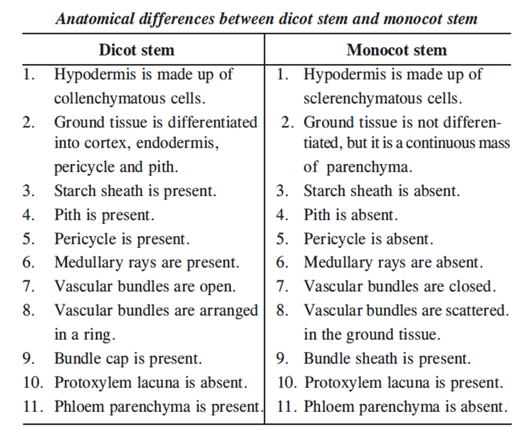
ISC 12>MULTICELLULARITY IN PLANTS>1. PLANT ANATOMY>Anatomical differences

SCOPE OF SYLLABUS

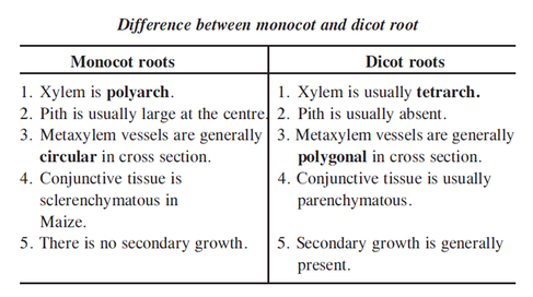
  • Anatomical differences between dicot and monocot root, stem and leaf must be taught for better understanding. 
  • Basic idea of how secondary growth takes place and formation of annual rings; structural and functional differences between heartwood and sapwood.(NEXT)

Click on the links below for plant tissues and secondary growth
Plant tissues (types of vascular bundles)                                                           Basic idea of secondary growth


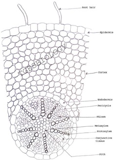
1. ANATOMY OF DICOT ROOT  (PRIMARY GROWTH)
DRAW THE CELLULAR DIAGRAM
Picture

DRAW THE SCHEMATIC DIAGRAM

Picture
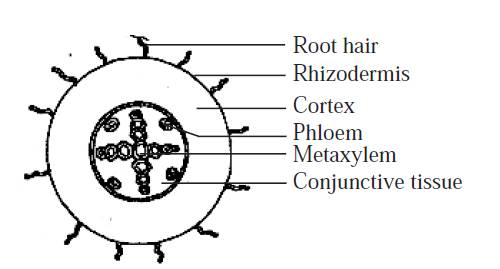
Primary structure of dicot root 
The transverse section of the dicot root shows the following plan of arrangement of tissues from the periphery to the centre.
  1. Rhizodermis or epiblema :
  • The outermost layer made up o single layer of parenchymatous cells without intercellular spaces.Stomata and cuticle are absent. 
  • Root hairs are always single celled. 
2.     Cortex:
  • Cortex consists of  oval or rounded loosely arranged parenchymatous cells. 
  • These cells may store food reserves.
3.     Endodermis 
  • It is made up of single layer of barrel shaped parenchymatous cells.
  • The radial and the inner tangential walls of endodermal cells are thickened with suberin.  These thickenings are known as casparian strips.
  • But these casparian strips are absent in the endodermal cells (Passage cells) which are located opposite to the protoxylem elements. 
4.    Stele:  All the tissues present inside endodermis comprise the stele.
       A.    Pericycle : Pericycle is generally a single layer of parenchymatous cells found inner to the endodermis. Lateral roots originate from the pericycle. 
      B.   Vascular system
  •  Vascular tissues are in radial arrangement. 
  • The tissue by which xylem and phloem are separated is called conjunctive tissue.
  • Xylem shoes exarch and  tetrarch condition . 
  • Metaxylem vessels are generally polygonal in shape
5. Pith: Usually absent
Root: Labeling exercise  

Images of Dicot root

Picture
http://pixgood.com/dicot-root-under-microscope.html
Part showing the vascular tissues
Picture
http://dulukbio.info/DulukBiology/Plants_files/ Ranunculus%20Root.jpg
Picture
http://faculty.hcc-nd.edu/lunfried/WWW(S2005)/BIOL-101(S2005)/Plant-Structure/Str-Photo/35-15a-XSDicotRoot.jpg

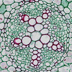
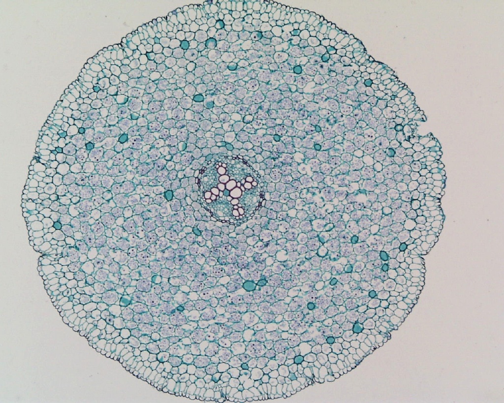
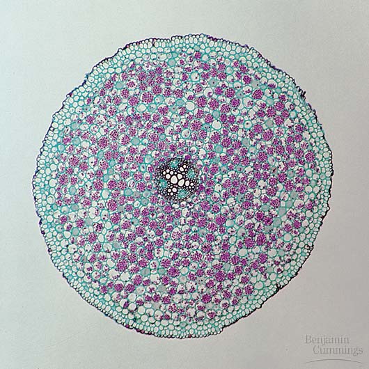
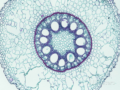
2. ANATOMY OF MONOCOT ROOT 

Primary structure of monocotyledonous root - Maize root 

 1.  Rhizodermis or epiblema
  • The outermost layer of parenchymatous cells without intercellular spaces.Stomata and cuticle are absent. 
  • Root hairs are always single celled.
2.  Cortex
  • The cortex is homogenous, consists of oval or rounded loosely arranged parenchymatous cells.  
  •  The function of cortical cells is storage.
3.  Endodermis 
  • It is made up of single layer of barrel shaped parenchymatous cells.
  • The radial and the inner tangential walls of endodermal cells are thickened with suberin. These thickenings are known as casparian strips.
  • But these casparian strips are absent in the endodermal cells (Passage cells) which are located opposite to the protoxylem elements. 
4.  Stele:  All the tissues present inside endodermis comprise the stele.
       A.    Pericycle : A single layer of parenchymatous cells found inner to the endodermis. Lateral roots originate from the pericycle. 
      B.   Vascular system
  •  Vascular tissues are in radial arrangement. 
  • Xylem and phloem are separated by sclerenchymatous conjunctive tissue.
  • Xylem shows exarch and polyarch condition. 
  • Metaxylem vessels are generally circular in shape.
5. Pith: 
  • The central portion is occupied by a large pith consist of thin walled parenchyma cells with intercellular spaces.
  •  These cells are filled with abundant starch grains.

DRAW THE  CELLULAR DIAGRAM 

Picture

DRAW THE SCHEMATIC DIAGRAM

Picture

Images of monocot root

Picture
http://biology.uco.edu/bidlack/botany/botanypics/ plant%20cells/zea%20root%201.jpg
Picture
http://imgc.allpostersimages.com/images/P-473-488-90/64/6474/BPQH100Z/posters/biodisc-cross-section-of-a-corn-primary-root-zea-mays-a-monocot-lm-x12.jpg
Picture
http://www.microbehunter.com/wp/wp-content/uploads/2009/maize_root1.jpg
Picture

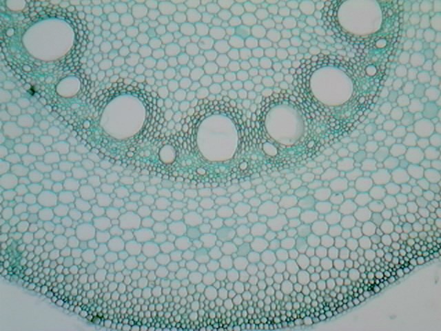
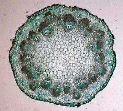
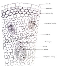
3. ANATOMY OF DICOT STEM (PRIMARY GROWTH)
DRAW THE DIAGRAM 
Picture

DRAW THE SCHEMATIC DIAGRAM

Picture

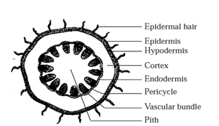
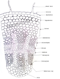
Primary structure of dicotyledonous stem - Sunflower stem 

1.  Epidermis
  • It is a protective outermost single layer of parenchymatous cells without intercellular spaces.
  • The outer walls of the epidermal cells have a layer called cuticle and multicellular hairs (trichomes)
2.  Cortex
  • Below the epidermis  cortex is differentiated into few layers of collenchyma cells that make hypodermis gives mechanical strength to the stem.
  • A few layers of chlorenchyma cells are present with conspicuous intercellular spaces. . Some resin ducts also occur here. 
  • The third zone is made up of parenchyma cells. These cells store food materials.
 3.  Endodermis: (Starch sheath)
  • The cells of this layer are barrel shaped arranged compactly without intercellular spaces. 
  • Due to abundant starch grains in these cells, this layer is also known as starch sheath. 
3.  Stele
  • It consists of pericycle, vascular bundles and pith. 
A)  Pericycle (Bundle cap)
  • Pericycle occurs between the endodermis and vascular bundles in the form of a few layers of sclerenchyma cells patches outside the phloem in each vascular bundle. (Bundle cap)
B)  Vascular bundles
  • In dicot stem, vascular bundles are arranged in a ring around the pith.  
  • Each vascular bundle is conjoint, collateral, open and endarch.
C)  Pith
  • The large central portion called pith composed of parenchyma cells with intercellular spaces.  
  • The extension of pith between vascular bundles are called as pith ray or medullary rays.
  •  Function of the pith is storage of food.
Dicot stem anatomy: Labeling exercise 

Images for dicot stem

Picture
http://www.lima.ohio-state.edu/academics/biology/images/dicotstem.jpg
Picture
http://29.media.tumblr.com/tumblr_lj9kepk68y1qiapgro1_500.jpg
Picture
http://www.bio.miami.edu/dana/pix/dicot_stem.jpg
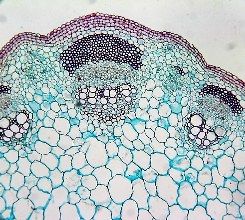
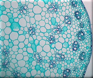
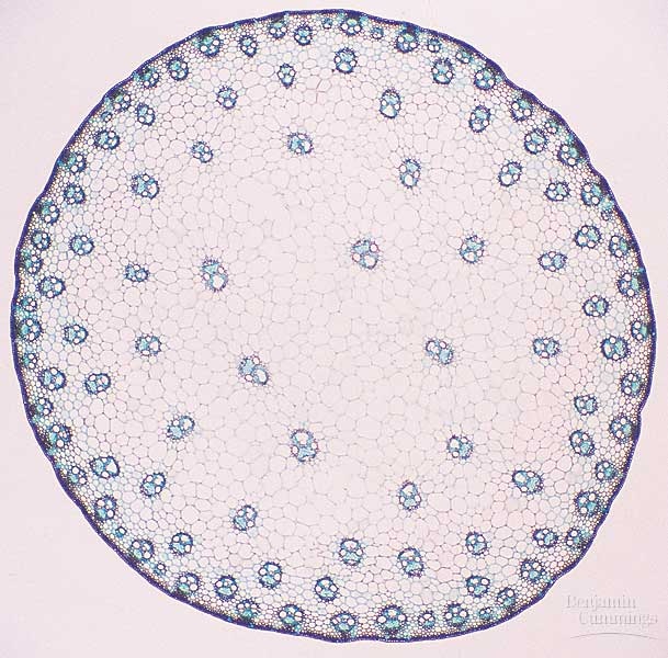
4. ANATOMY OF MONOCOT STEM

Primary structure of monocot stem - Maize stem
1.  Epidermis
  • It is the outermost layer made up of single layer of tightly packed parenchymatous cells with thick cuticle. 
  • There are no epidermal outgrowths.
2.   Hypodermis
  • A few layer of sclerenchymatous cells lying below the epidermis constitute the hypodermis, gives mechanical strength to the plant.
3.  Ground tissue
  •  It is not differentiated into cortex, endodermis, pericycle and pith. 
  • The ground tissue is represented by several layers of loosely arranged parenchyma cells enclosing prominent intercellular spaces.
  •  The ground tissue is meant for storage of food. 
4.  Vascular bundles
  • Vascular bundles are scattered in the parenchymatous ground tissue.
  • Vascular bundles are numerous, small and closely arranged in the peripheral portion.
  • Towards the centre, the bundles are comparatively large in size and loosely arranged. 
  • Each vascular bundle is surrounded by a sheath of sclerenchymatous fibres called bundle sheath.
  • The vascular bundles are conjoint, collateral, endarch and closed.  
Phloem: 
  • The phloem in the monocot stem consists of sieve tubes and companion cells. 
  • Phloem parenchyma and phloem fibres are absent.
Xylem :
  • The two metaxylem vessels are located at the upper two arms and one or two protoxylem vessels at the base. (Y shaped)
  •  In a mature bundle, the lowest protoxylem disintegrates and forms a cavity known as protoxylem lacuna.
DRAW THE DIAGRAM 
Picture

DRAW THE SCHEMATIC DIAGRAM

Picture

Picture
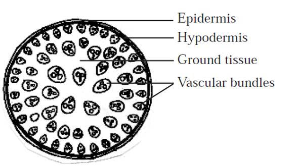
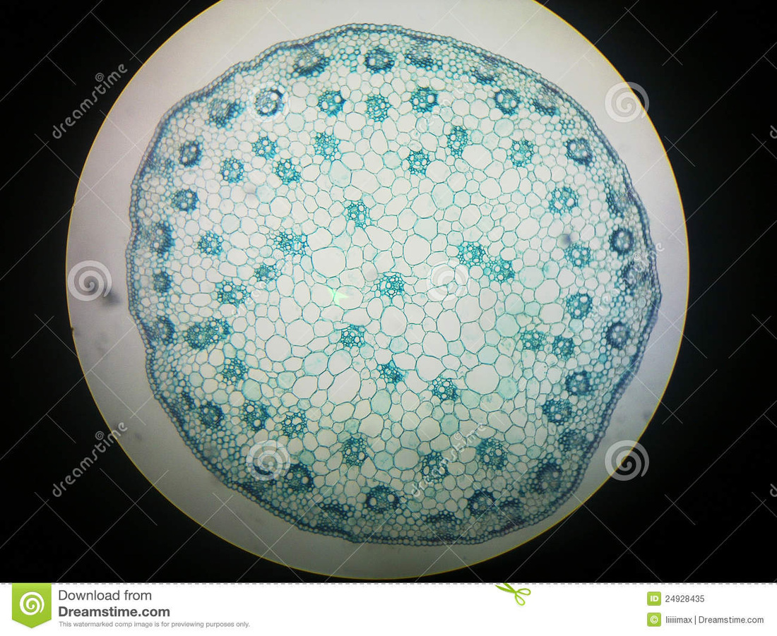
http://thumbs.dreamstime.com/z/cross-section-corn-stem-24928435.jpg
Picture
http://www.microscopy-uk.org.uk/mag/imgaug03/brunel6.jpg
Picture
http://faculty.hcc-nd.edu/lunfried/WWW(S2005)/BIOL-101(S2005)/Plant-Structure/Str-Photo/35-18b-MonocotStem.jpg
Picture

Dicot vascular bundle

Picture

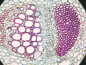
Monocot vascular bundle

Picture

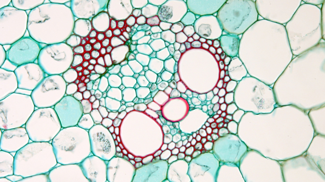
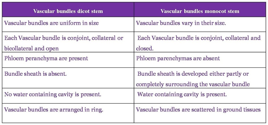
Differences between monocot and dicot vascular bundles

Picture

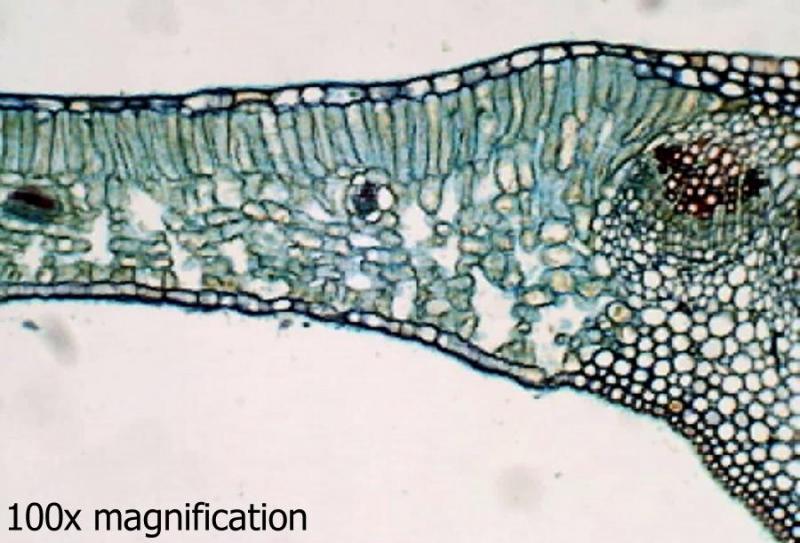
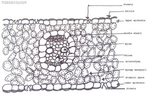
5. ANATOMY OF DICOT LEAF (Dorsiventral leaf)                 
Picture
Picture
http://mail.gibraltar.k12.wi.us:8100/~jdickson/ AP_Biology/Lab19PlantAnatomy/images/lilac_leaf_cs.jpg
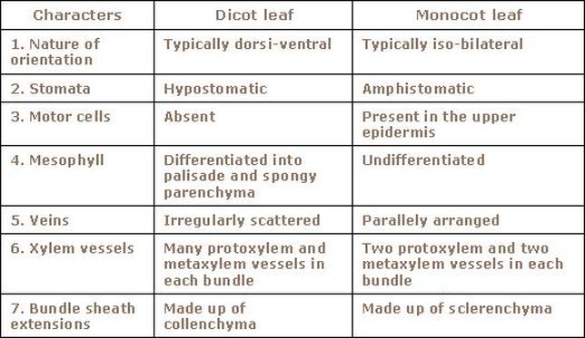
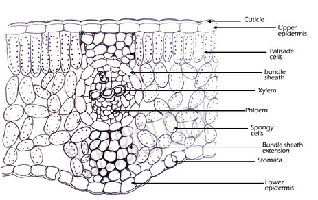
A transverse section through the mid rib region of a typical dorsiventral leaf reveals the following structure. 
1. Epidermis
  • Both upper and lower epidermis are usually made up of a single layer of cells that are closely packed.
  • The cuticle on the upper epidermis is thicker than that of lower epidermis.
  • Stomata are more in number on the lower epidermis than on the upper epidermis.
  • The main function of the epidermis is to give protection to mesophyll. The cuticle helps to check transpiration. Stomata are used for transpiration and gas exchange.
2 . Mesophyll : 
  • The entire tissue between the upper and lower epidermis is called the mesophyll, differentiated into palisade and spongy parenchyma.
A)   Palisade parenchyma 
  • Below the epidermis, vertically elongated cylindrical cells in one or more layers without intercellular spaces form palisade parenchyma.
  • Palisade parenchyma cells contain more chloroplasts than the spongy parenchyma cells. The function of palisade parenchyma is photosynthesis. 
B)   Spongy parenchyma
  • Below palisade parenchyma towards lower epidermis, irregularly shaped, loosely arranged cells with numerous air spaces form spongy parenchyma. 
  • Spongy cells facilitate the exchange of gases with the help of air spaces.
  • The air space that is found next to the stoma is called respiratorycavity or sub-stomatal cavity
3)   Vascular tissues (in the veins)
  • Vascular bundles are conjoint, collateral and closed. Xylem is present towards the upper epidermis, while the phloem towards the lower epidermis.
  • Vascular bundles are surrounded by a compact layer of parenchymatous cells called bundle sheath or border parenchyma. 
  • Protoxylem vessels are present towards the upper epidermis. 
  • Phloem consists of sieve tubes, companion cells and phloem parenchyma. Phloem fibres are absent. 
  • Xylem consists of vessels and xylem parenchyma. Tracheids and xylem fibres are absent.
ONLINE ASSESSMENT

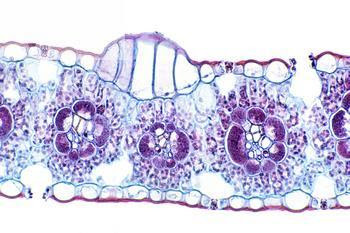
6. ANATOMY OF MONOCOT LEAF  (Isobilateral leaf)
Picture
Picture
http://3.bp.blogspot.com/_d1nJkEYl80o/RljvnlmKPkI/ AAAAAAAAA3c/Bm6E7UgjyOs/s400/55544.jpg
A transverse section passing through the midrib region of an iso-bilateral leaf (Maize) reveals the following structure.
1.  Epidermis 
  • Both upper and lower epidermis are usually made up of a single layer of cells that are closely packed.
  • Cuticle and trichomes are present in both the layers. 
  • Stomata are equal in number on both layers.  This condition is described as amphistomatic.
  • A few cells in the upper epidermis are enlarged to form motor cells called bulliform cells.Helps in rolling and unrolling.
2.  Mesophyll
  • It is ground tissue that occurs between the two epidermal layers not differentiated into palisade and spongy parenchyma.
  • It is composed of many layers of loosely arranged, spherical or oval chlorenchyma cells. Has few intercellular spaces.
3.  Vascular bundles
  • Each vascular bundle is surrounded by a bundle sheath composed of a single layer of compactly arranged cells. 
  • Xylem is found towards upper epidermis and phloem is towards lower epidermis. The vascular bundle is described as conjoint and collateral with endarch xylem.
  • The oldest and the largest vascular bundle is found in the centre. It is known as midrib vein. The bundle sheath of the midrib vein is connected to the upper and lower epidermal layers by sclerenchyma cells representing bundle sheath extensions or hypodermal sclerenchyma.

Difference between dicot and monocot leaf
Picture

Plant tissues                                                                                Basic idea of secondary growth

Any suggestions to improve the website content are welcome, and feedback that points out errors and ambiguities will be appreciated................ Contact:Gajendra Khandelwal(mandawara8@gmail.com), Deepthi.Uthaman (duthaman12@gmail.com)