ISC 11>DIVERSITY OF LIFE>2-KINGDOM PROTISTA
SCOPE OF SYLLABUS
- General characteristics of Kingdom Protista -
- Characteristics and examples of subgroups:
- Protozoans to be studied under rhizopods, flagellates, ciliates and sporozoans with brief characteristics and common examples of each.
CLASS PRESENTATION ANIMATIONS
|
|
| ||||||||
CHARACTERISTICS OF PROTISTA
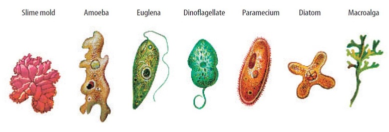
- Protistans are unicellular eukaryotes.
- They possess membrane-bound distinct organelles like nucleus with nuclear membrane and nucleoli, mitochondria, chloroplasts (in photosynthetic protists), Golgi bodies and various types of microbodies.
- Photosynthetic forms have a distinct cell wall. Some protists (predatory protozoans) have special organelles for the intake of food and elimination of waste.
1. Mode of locomotion
- Locomotion by Pseudopodia: The locomotion by pseudopodia is a slow (0.2—3.0 µm/sec) and creeping type and is called amoeboid movement. It takes place with the help of protoplasmic outgrowths called pseudopodia. This type of locomotion is seen in sarcodines and slime moulds. Pseudopodia are of four types:
- (a) Lobopodia are lobe-like pseudopodia with broad and blunt ends. These are present in Amoeba.
- (b) Filopodia are fine and thread-like tapering pseudopodia. These are found in Euglypha.
- (c) Axopodia are hard and stiff with hard axial ifiament. These are found in Actinophrys.
- (d) Reticulopodia are long and branching pseudopodia that form a network as in Globigerina.
- Some protists move with the help of long, whip-like and fine structures, flagella.
- There may be one flagellum as in Euglena or two as in chlamydomonas or more than two.
- Flagellar locomotion occurs in dinoflagellates (Gonyaulax and Gymnodinium), euglenoids (Euglena) and zooflagellates (Leishmania, Trypanosoma).
2. Mode of nutrition
Following modes of nutrition are found in protists:
- Photosynthetic or Holophytic Nutrition: Some protists synthesise their food from CO2 and water utilising sun’s energy in the presence of photosynthetic pigments like chlorophyll. Examples: Dinoflagellates, Diatoms and Euglenoids.
- Holozoic or Zootrophic Nutrition: In this case, food is ingested like animals. Examples: Amoeba, Paramecium
- Saprobic Nutrition: In this mode of nutrition, organisms secrete enzymes in the surrounding medium where organic matter is converted into simple molecules, which are absorbed through the body surface of the organism. Example: Slime moulds.
- Parasitic Nutrition: Parasitic protists obtain readymade food from other organisms. The organism which provides food to the parasite is called host. Examples: Plasmodium, Entamoeba, Trypanosoma, Giardia.
- Myxotrophic Nutrition: In this case, the protistans show mixed type of nutrition, i.e., autotrophic and heterotrophic. Example: Euglena.
- Symbiotic: Zooflagellates like Trichonympha in termites and Lophomonas in woodroaches live as symbionts in some other organisms and by secreting cellulose-digesting enzymes convert cellulose into glucose.
3. Reproduction
Protists have great reproductive potential and reproduce both by asexual and sexual methods.
- Asexual Reproduction: Under favourable conditions, protists can reproduce several times a day leading to population explosion. Since asexual reproduction involves only one parent, the offspring produced by this method are all alike genetically to the parent and are termed as clones. Asexual reproduction may occur in the following ways:
- Binary Fission: It is the division of one parent cell into two daughter cells. It occurs under favourable conditions and takes place by the division of nucleus followed by the division of cytoplasm. After binary fission, each daughter cell becomes an independent organism. Binary fission may be irregular as in Amoeba, transverse as in Paramecium or longitudinal as in Euglena.
- Multiple Fission: It is the division of the parent organism into several daughter cells, each containing one nucleus. Each daughter cell grows into an independent organism as in Amoeba and Plasmodium vivax, etc.
- Budding: In some protists, a small outgrowth develops from the parent body, enlarges in size and finally separates and develops into a new individual. This is called budding. Example: Arcella.
- Cyst Formation: Many protists, at the approach of unfavourable climatic conditions become metabolically inert and form a thick and resistant covering around them. This is called cyst. On the approach of favourable conditions, cyst imbibes water, ruptures and an active organism comes out. Many protozoan diseases like amoebiasis, are spread through cysts, e.g., Entamoeba.
- Plasmotomy: It is the division of multinucleate protist into two or more multinucleate offspring. Example: Opalina, Pelomyxa.
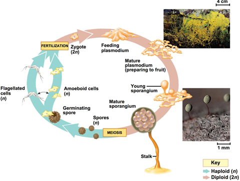
- Spore Formation: In some protists, spores or propagules are formed. They have a protective cover to withstand unfavourable conditions. On germination, each spore gives rise to a new individual. Example: Slime moulds.
- Sexual reproduction involves fusion of two haploid (n) cells, the gametes. The process of fusion is called fertilisation or syngamy. It produces a diploid (2n) cell called zygote.
- It undergoes mitotic divisions to give rise a number of diploid daughter cells or undergoes meiotic division to form haploid daughter cells.
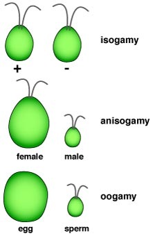
- Syngamy occurs through
- (I) Isogamy (Two fusing gametes that are structurally and functionally similar, e.g., Monocystis);
- (ii) Anisogamy (Two fusing gametes are dissimilar, e.g., Ceratium) and
- (iii) Oogamy (Large non-motile female gamete is fertilised with smaller motile male gamete, e.g., Plasmodium).
- The process of meiosis is an essential step in sexual reproduction because it reduces the chromosome number to half in gametes.
- After fertilisation, the diploid number of chromosomes (2n) is restored and is maintained constantly in a species.
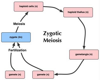
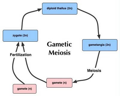
Life cycle in protista: In general, two types of life cycles are found in protists: (EXTRA INFORMATION)
|
1. Life Cycle with Zygotic Meiosis:
|
2. Life Cycle with Gametic Meiosis:
|
|
|
4. Classification
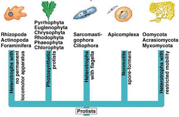
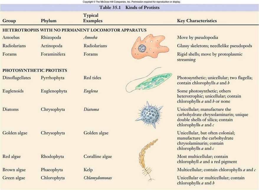
Major groups of protists
A. PHOTOSYNTHETIC PROTISTS
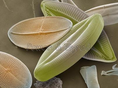
1. CHRYSOPHYTES : Diatoms and golden algae
This group includes diatoms and golden algae (desmids). Chrysophyta is the golden algae. Diatoms and golden algae are grouped together because they both produce a unique carbohydrate called chrysolaminarin.
This group includes diatoms and golden algae (desmids). Chrysophyta is the golden algae. Diatoms and golden algae are grouped together because they both produce a unique carbohydrate called chrysolaminarin.
| diatomsmoving-v1.swf |
- Habitat: -They are found in fresh water as well as in marine environments. They are microscopic and float passively in water currents (plankton).
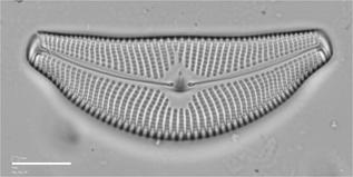
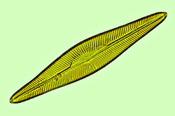
- Forms : Unicellular organisms with unique double shells made of silica, which are often strikingly and characteristically marked. The shells of diatoms are like small boxes with lids, one half of the shell fitting inside the other. There are two major groups of diatoms, one with radial symmetry (like a wheel) and the other with bilateral (two sided) symmetry.
- Nutrition : Photosynthetic
- Pigments : Their chloroplasts with chlorophylls a and c, fucoxanthin, xanthophylls.
- Locomotion : A flagella is absent except in the reproductive stages. The cells may exhibit gliding type of movement with the help of mucilage secretion.
- Reproduction :
- Asexual : Diatom shells are rigid, and the organisms reproduce asexually by separating the two halves of the shell, each half then regenerating another half shell within it. Because of this mode of reproduction, there is a tendency for the shells, and consequently the individual diatoms, to get smaller and smaller with each asexual reproduction. When the resulting individuals have diminished to about 30% of their original size, one may slip out of its shell, grow to full size, and regenerate a full-sized pair of new shells.
- Sexual : Individual diatoms are diploid. Meiosis occurs more frequently under conditions of starvation. Some marine diatoms produce numerous sperm and others a single egg. If fusion occurs, the resulting zygote regenerates a full-sized individual.
- Significance :
- The walls are embedded with silica and thus the walls are indestructible. Thus, diatoms have left behind large amount of cell wall deposits in their habitat; this accumulation over billions of years is referred to as ‘diatomaceous earth’.
- Being gritty this soil is used in polishing, filtration of oils and syrups.
- Diatoms are the chief ‘producers’ in the oceans.
- Example : Cymbella, Navicula, Melosira
|
Cymbella sp.
|
Navicula sp
|
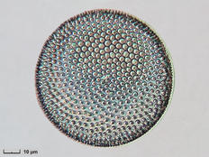
Coscinodiscus sp.
|
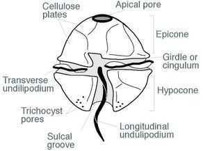
2. PYRROPHYTA: The Dinoflagellates
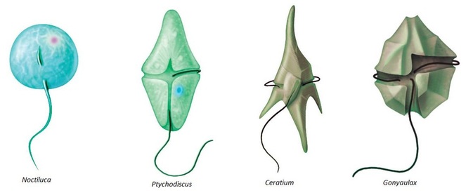
- Habitat: A majority of the dinoflagellates are marine, and they are often abundant in the plankton, but some occur in fresh water.
- Forms : Unicellular
- The cell wall has stiff cellulose plates on the outer surface.
- Most of them have two flagella; one lies longitudinally and the other transversely in a furrow between the wall plates.
- Nutrition : Photosynthetic and reserve food is starch and oil.
- Pigments : They appear yellow, green, brown, blue or red depending on the main pigments present in their cells. Most have chlorophylls a and c, in addition to carotenoids.
- Locomotion:
- Most of have two flagella. The flagella are usually located within grooves, one encircling the body like a belt, and the other perpendicular to it. By beating in their respective grooves, these flagella cause the dinoflagellate to rotate like a top as it moves.
- Significance:
- Very often, red dianoflagellates (Example: Gonyaulax) undergo such rapid multiplication that they make the sea appear red (red tides).
- Toxins released by such large numbers may even kill other marine animals such as fishes.
- More recently, a particularly dangerous toxic dinoflagellate called Pfiesteria piscicida is reported to be a carnivorous, ambush predator. During blooms, it stuns fish with its toxin and then feeds on the prey’s body fluids.
- Example :
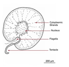
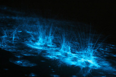
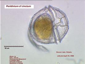
- Some planktonic dinoflagellates are luminous and contribute to the twinkling or flashing effects that we sometimes see in the sea at night, especially in the tropics. Noctiluca, Peridinium and Gonyaulax
|
Noctiluca
|
Peridinium
|
Ceratium
|
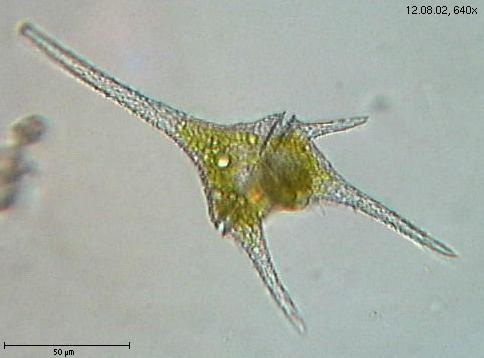
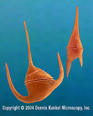
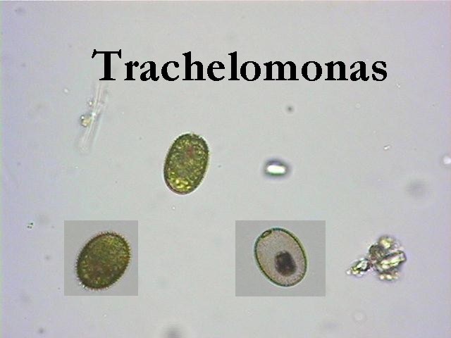
3. EUGLENOPHYTA: EUGLENOIDS
- Habitat: Majority of them are fresh water organisms found in stagnant water.
- Forms :
- The members of this phylum clearly illustrate the impossibility of distinguishing “plants” from “animals” among the protists.
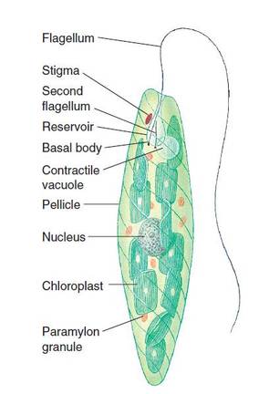
- They devoid of cellulosic cell wall. Interlocking proteinaceous strips arranged in a helical pattern form a flexible structure called the pellicle, which lies within the cell membrane of the euglenoids. Because its pellicle is flexible, a euglenoid is able to change its shape.
- Nutrition: About a third of the approximately 40 genera of euglenoids have chloroplasts and are fully autotrophic; the others lack chloroplasts, ingest their food, and are heterotrophic. Some euglenoids with chloroplasts may become heterotrophic if the organisms are kept in the dark; the
- chloroplasts become small and nonfunctional. If they are put back in the light, they may become green within a few hours. Reserve food is in the form of paramylum bodies.
- Pigments : Cells of Euglena contain numerous small chloroplasts. These chloroplasts, like those of the green algae and plants, contain chlorophylls a and b, together with carotenoids.
- Locomotion : In Euglena , the genus for which the phylum is named, two flagella are attached at the base of a flask-shaped opening called the reservoir, which is located at the anterior end of the cell. One of the flagella is long and has a row of very fine, short, hairlike projections along one side. A second, shorter flagellum is located within the reservoir but does not emerge from it.
- Eye spot: The stigma, an organ that also occurs in the green algae (phylum Chlorophyta), is light-sensitive and aids these photosynthetic organisms to move toward light.
- Contractile vacuoles: Collect excess water from all parts of the organism and empty it into the reservoir, which apparently helps regulate the osmotic pressure within the organism.
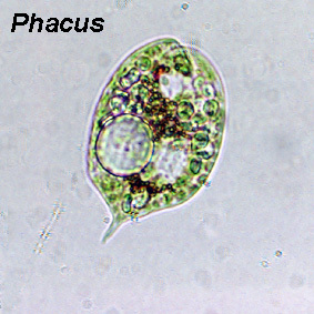
- Example : Eugleana, Phacus, Astasia, Trachelomonas
|
|
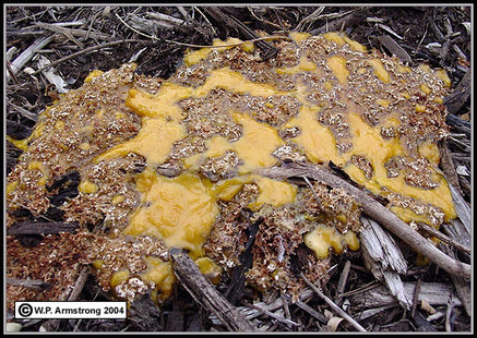
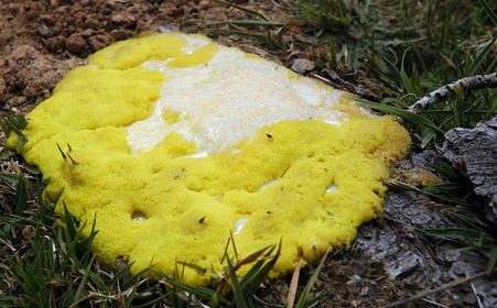
B. CONSUMER DECOMPOSER PROTISTS: SLIME MOLDS
Resemble fungi in appearance and life-style, but their cellular organization, reproduction, and life cycles are more closely related to protists.
|
| ||
|
CHARACTERISTICS OF SLIME MOULDS
1. They are seen during rainy season, in cool, shady places.
2. Cell wall is present only in spores. 3. Mostly heterotrophic, some are saprotrophic or parasitic. 4. Exhibit amoeboid movement, some are swarmers that move with the flagella. 5. Reproduction is both asexual and sexual. 6. They occur in a wide variety of colours. |
|
|
FEATURES LIKE PLANTS
1. They show formation of spores in sporangium, with walls made up of cellulose.
2. Show saprophytic mode of nutrition. 3. Reproduction is isogamous. |
FEATURES LIKE ANIMALS
1. No chlorophyll, vegetative body is coenocytic and without cell wall.
2. Shows characteristic amoeboid movement. 3. Holozoic mode of nutrition. |
DIFFERENCES WITH FUNGI
The slime moulds look like fungi- however they are different from Fungi in the following ways-
1. They can move whereas fungi are stationary.
2. They ingest food whereas fungi absorb food.
3. The cell wall is made up of cellulose and chitin.
1. They can move whereas fungi are stationary.
2. They ingest food whereas fungi absorb food.
3. The cell wall is made up of cellulose and chitin.
CLASSIFICATION
There are two groups of slime moulds-
A. Acellular or plasmodial slime moulds- class Myxomycetes.
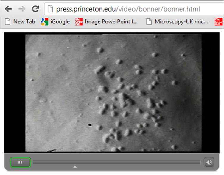
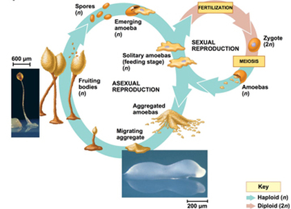
B. Cellular slime moulds- class Acrasiomycetes.
A. Acellular or plasmodial slime moulds- class Myxomycetes.
B. Cellular slime moulds- class Acrasiomycetes.
|
Acellular slime mould
|
Cellular slime mould
|
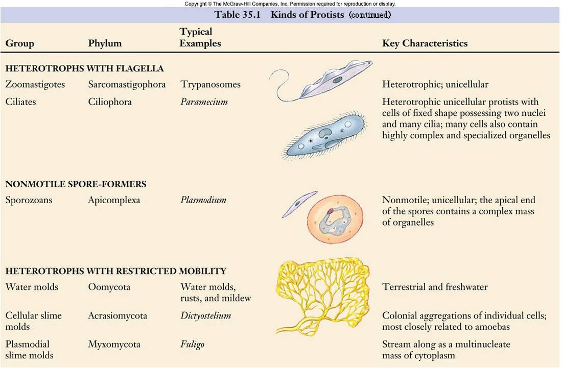
C. PROTOZOAN PROTISTA
General characteristic
- Structure: Unicellular body consists of a mass of protoplasm differentiated into cytoplasm and one or more nuclei.
- Body is protected by a firm pellicle, rigid wall or silicious shell.
- Locomotion: It may take place by blunt, finger-like processes, the pseudopodia, or by long whip-like flagella or by hair-like processes, the cilia.
- Nutrition: Nutrition is mainly holozoic. Some forms have saprobic nutrition.
- Respiration: Exchange of gases takes place through the general body surface.
- Excretion: No definite organ for excretion. It takes place through general body surface.
- Osmoregulation: Freshwater forms have one or more contractile vacuoles to get rid of excess water.
Major subgroups
- Amoeboid protozoans:
- Flagellated protozoans:
- Ciliated protozoans:
- Sporozoans:
1. Amoeboid protozoans:
1. Amoeba proteus EXTRA INFORMATION
- Habitat: These organisms live in fresh water, sea water or moist soil.
- Nutrition: Heterotrophic They move and capture their prey by putting out pseudopodia (false feet) as in Amoeba. Some of them such as Entamoeba are parasites.
- Locomotion: By pseudopodia
- Reproduction: Asexual reproduction by binary fission and sexual reproduction if present is by syngamy.
- Marine forms have silica shells on their surface.
1. Amoeba proteus EXTRA INFORMATION
- It was discovered by Russel von Rosenhoff in 1755. Amoeba is found in freshwater. Its pseudopodia are of labopodia type. A contractile vacuole is present for osmoregulation. Cytoplasm is differentiated into endoplasm and ectoplasm. Endoplasm is further differentiated into plasmagel and plasmasol. The entire body is covered with plasmalemma. Nutrition is holozoic. Ingestion takes place by following methods:
- 1. Import, the passive food-ingestion.
- 2. Invagination by forming an invagination tube to take food particles.
- 3. Circumfluence for less active food particles.
- 4. Circtmivallation is for active preys.
- Digestion occurs inside food vacuole. The contents of food vacuole become first acidic and then alkaline. Sol-gel theory for amoeboid locomotion was given by Hyman which was later on supported by Pantin and Mast.
- Asexual reproduction occurs by binary fission and sporulation (multiple fission). Cyst formation takes place in unfavourable conditions. Sexual reproduction is absent.
- It was discovered by Lamble in 1859 and Losch gave its pathogenic nature in 1875. Life cycle of Entamoeba is monogenetic, i.e., it completes its life cycle in a single host (i.e., man). Entamoeba resides in the upper part of the human large intestine and causes amoebiasis (amoebic dysentery).
- Symptoms of this disease are abdominal pain, repeated motions with blood and stool. Parasite has only one pseudopodium. Contractile vacuole is absent. It feeds on red blood corpuscles by damaging the wall of large intestine and produces ulcers.
- E. histolytica occurs in two forms — (i) magna (trophozoite), the pathogenic form found in the mucosa and submucosa of intestine forming ulcers and (ii) minuta (nonpathogenic form) found in the lumen of the intestine. A mature cyst is called quadrinucleate cyst. It has four nuclei and two chromatoid bodies. The quadrinucleate cyst is the infective stage of Entamoeba. On entring the intestine of new host, the single cyst of E. histoiytica produces eight amoebae.
2. Flagellated protozoans
- The members of this group are either free-living or parasitic.
- They have flagella.
- The parasitic forms cause diseases such as sleeping sickness.
- Examples: Human parasites like Trypanosoma, Trichomonas, Leishmania, Giardia and gut parasites of insects like Trichonympha, Lophomonas, etc.
1. Trypanosoma (EXTRA INFORMATION) JUST REMEMBER THE EXAMPLES
- Trypanosoma gambiense: It is found in the central and the western parts of Africa. It causes African sleeping sickness in man. It is an endoparasite living in the blood and the cerebrospinal fluid of man. The parasite is transmitted by the bite of tse-tse fly, Glossina palpalis. The disease appears when the parasite enters the cerebrospinal fluid. It makes the patient lethargic and unconscious, therefore, the disease is called sleeping sickness.
- Trypanosoma rhodesiense: It causes Rhodesian sleeping sickness or Rhodesian trypanosomiasis. The parasite is transmitted by the bite of tse-tse fly (Glossina palpalis and Glossina morsitans). After the infection, the parasite initially and later on enters the blood.
- Trypanosoma cruzi: It causes Chagas disease. The symptoms of the disease are fever, diarrhoea, anaemia and enlargement of lymphoid glands. Infection takes place by contamination of wounds with faeces of triatomid bugs.
- Leishmania donovani: It causes kala-azar. This disease is common in East Asian countries, India and parts of Africa and America. The fever is accompanied with anaemia, enlargement of liver and spleen. Dogs and cats are reservoir host. The parasite is transmitted by a sand fly, Phiebotomus argentipes and other species. The parasite lives in the cells of liver, spleen, lymph glands and bone marrow.
- Leishmania tropica: It causes oriental sore in humans. The disease is characterised by cutaneous sores on hands, feet and face. The disease spreads by sand flies. The parasite lives in the endothelial cells of skin capillaries.
- Leishmania brasilliensis: It causes espunda disease in man. The disease is characterised by lesions on skin and mucous membrane of nose, mouth, pharynx and sometimes vagina. The disease is transmitted by sand flies.
- It causes giardiasis in human beings. It is characterised by epigastric pain, abdominal discomfort, diarrhoea, headache and fever. Transmission occurs by taking cysts of the parasite with contaminated food and water.
- It inhabits vagina of woman and causes leucorrhoea. It is characterised by burning sensation, itching and frothing discharge. Its transmission is by sexual intercourse. In male, infection of urethra and prostate gland is common.
- It occurs as a symbiont in the intestine of termites. It secretes cellulose digesting enzymes which convert cellulose into glucose. The digested food is shared by the symbiont and termite.
- It occurs as a symbiont in the intestine of termites and wood roaches. It secretes enzymes for the digestion of cellulose. The digested food is shared by both the host and the parasite.
3. Ciliated protozoans:
- These are aquatic,
- Nutrition is holozoic.
- They have a cavity (gullet) that opens to the outside of the cell surface. The coordinated movement of rows of cilia causes the water laden with food to be steered into the gullet. Cell organelles like cytopharynx, neuromotor system, contractile vacuoles and cytopyge are present.
- Trichocysts, the organelles of defence, are present. These are ejected out with force to stun the prey.
- Nuclei are of two types: a large macronucleus that controls the vegetative activities and one or more micronuclei which take part in sexual reproduction.
- Asexual reproduction by transverse binary fission.
- Sexual reproduction by means of conjugation.
- Actively moving organisms because of the presence of thousands of cilia.
- Example: Paramoecium
1. Paramecium EXTRA INFORMATION (NOTE: Just remember the names)
- Paramecium, the slipper-shaped animalcule, is found in freshwater ponds and slow streams containing decaying organic matter.
- It is an active swimmer with holozoic mode of nutrition.
- It is 0.25 mm long. It bears cilia all over the body.
- The free movement is brought about by beating of cilia in a coordinated manner.
- Reproduction: Paramecium reproduces asexually by binary fission and sexually by conjugation. It encysts in unfavorable conditions.
- In conjugation, two individuals come in contact with each other by ventral side. The diploid micronucleus of each individual undergoes meiosis to form haploid gametic nuclei. These are exchanged and the two individuals separate. The exchange of genetically different nuclei in Paramecium is essential.
- Balantidium coli lives as an endoparasite in large intestine of man. It causes ciliary dysentery. Transmission of parasite is through cysts in contaminated food and water.
- Treatment: Aureomycin, Terramycin and Carbarsone are ideal for curing the ciliary dysentery caused by Balantidium.
4. Sporozoans:
- Locomotion: Sporozoans have no physical form of movement. However, they can be moved by the currents of the blood or other fliuds of their hosts.
- Nutrition: They are parasites. Sporozoans have special organelles that allow it to invade a host cell.
- Life cycle may include more than one host.
- Asexual reproduction takes place by binary fission and sexual reproduction if present, by syngamy followed by the formation of haploid spores, the sporozoites.
- Examples: Plasmodium, Monocystis and Eimeria.
- The most notorious is Plasmodium (malarial parasite) which causes malaria which has a staggering effect on human population.
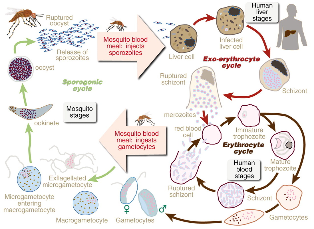
MALARIA PARASITE LIFE CYCLE:
1. Plasmodium (Malarial parasite) EXTRA INFORMATION
- Various species of Plasmodium cause different types of malaria in man. Its causal organism was discovered by Laveran in 1880.
- Sir Ronald Ross discovered that the disease spreads by mosquito bite.
- Life cycle of Plasmodium is digenetic, i.e., it is completed in two hosts:
- Female Anopheles mosquito (the primary host) and man (the intermediate or secondary host).
- The female Anopheles mosquito acts as vector or carrier of malarial parasite
- The female Anopheles mosquito carries the sporozoite stage of the protozoan. When it bites man, sporozoites enter the blood. From blood sporozoites enter the liver and change to schizonts. The schizonts produce cryptomerozoites which enter RBCs and form trophozoites. These become amoeboid and form pseudopodia. They grow in size and become schizonts. They by multiple fission produce merozoites. Some merozoites behave as gametes.
- When female Anopheles sucks blood, gametes pass into the gut. In the gut of mosquito sexual reproduction takes place by the fusion of gametes: The zygotes, so formed, produces needle- shaped sporozoites which enter the blood of man by mosquito bite.
|
|
SUMMARY VIDEO
