ISC 10>HUMAN ANATOMY AND PHYSIOLOGY> 7. EXCRETORY SYSTEM
Scope of syllabus
|
|
Why do we urinate more frequently in winters?
Our body has different methods of releasing liquids- by perspiration and by urination.
There are many metabolic reactions taking place in the body, these biochemical reactions produce useful substances as well as certain toxic substances. This can also cause a change in the body water content. It is essential to remove these toxic substances and maintain the water level of the body. The removal of these toxic metabolic waste is called excretion.
Waste products are in the form of carbon-di-oxide from respiration, bile pigment from breakdown of hemoglobin and the main form of waste is from the breakdown of excess amino acids. These can be in the form of ammonia, urea or uric acid. These are released out of the body in the urine.
Our body has different methods of releasing liquids- by perspiration and by urination.
- When the temperature is warmer we perspire more, hence frequency of urination is less.
- In cold winter months we perspire less, hence urinate more frequently.
- This is a part of maintaining homeostasis in the body.
- Urine is produced by the kidney, that is a part of the excretory system.
There are many metabolic reactions taking place in the body, these biochemical reactions produce useful substances as well as certain toxic substances. This can also cause a change in the body water content. It is essential to remove these toxic substances and maintain the water level of the body. The removal of these toxic metabolic waste is called excretion.
Waste products are in the form of carbon-di-oxide from respiration, bile pigment from breakdown of hemoglobin and the main form of waste is from the breakdown of excess amino acids. These can be in the form of ammonia, urea or uric acid. These are released out of the body in the urine.
Excretion |
Defecation |
Excretion is removal of nitrogenous wastes
|

Defecation is removal of undigested waste
|
Excretory organs and their products in humans
|
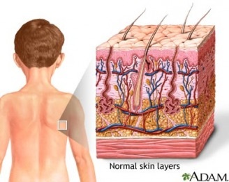
SKIN
|
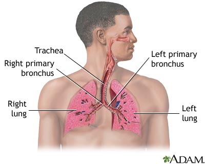
LUNGS
|
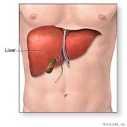
LIVER
|
KIDNEY
|
|
Sweat - contains urea and some salts
|
Carbon di oxide
|
Bile pigments
|
Urea
|
Extra information
Types of nitrogenous wastes
|
AMMONIA
Some living beings waste in the form of ammonia.
Ammonia is highly toxic. These organisms are known as ammoniotelic. Examples- Sponges, coelenterates and bony fishes. |
URIC ACID
Some living organisms release waste in the form of uric acid.
uric acid is least toxic. These organisms are called uricotelic. Examples- insects, reptiles and birds. |
UREA
Some living organisms release waste in the form of urea.
Urea is neutral and less toxic. These organisms are called ureotelic. Examples- desert mammals, cartilageous fishes and humans |
Few molluscs like Unio and Limnaea and Echinoderms excrete amino acids directly, these are called amminotelic animals
EXCRETORY SYSTEM IN HUMANS
|
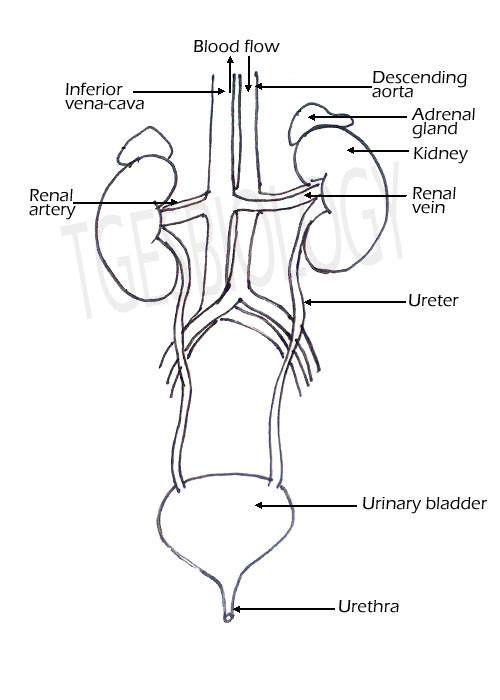
The excretory system or the urinary system consists of
|

http://upload.wikimedia.org/wikipedia/commons/1/18/Illu_urinary_system.jpg
|
DIAGRAM OF THE EXCRETORY SYSTEM
|
|
|
DRAW THE DIAGRAM
KIDNEY
|
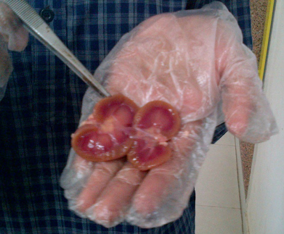
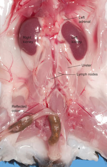
The kidneys are bean-shaped organs, about the size of a fist, with a smooth, reddish-brown surface. They have a convex shape on one side and a concave shape on the other side. In the middle of the concave side a notch is present through which structures enter and leave the kidney. This is called Hilum. 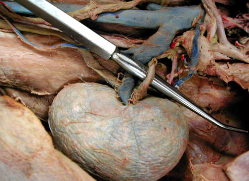
http://aahsanatomy.pbworks.com/f/1242221843/renals%5B1%5D.jpg
|
https://ars.els-cdn.com/content/image/3-s2.0-B9780128029008000038-f03-24-9780128029008.jpg
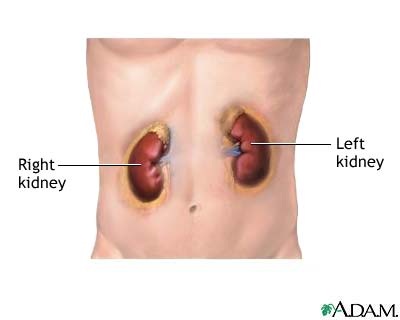
Location and position of the Kidney-
In the abdominal cavity below the stomach, on either side of the vertebral column. The right kidney is placed slightly lower than the left kidney as it is pushed down by the large right liver lobe. |
Blood vessels entering and leaving the kidney

|
The descending aorta branches to form the renal artery that enters the kidney, and the renal vein comes out of the kidney to join the inferior vena cava.
|
http://www.infobarrel.com/media/image/67136.jpg
|
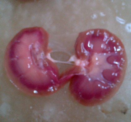
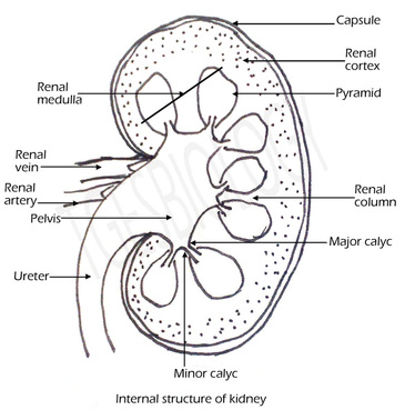
Internal structure of kidney-
Two distinct regions are seen-
The pyramids open into small tube like structures the minor calyc and two or three minor calyc opens into the a major calyc. The all the major calyces opens into the pelvis. The pelvis is the proximal end of the ureter that receives the urine from the calyces. |

http://training.seer.cancer.gov/images/anatomy/urinary/kidney.jpg
|
|
DRAW THE DIAGRAM
|
|
HOW THE KIDNEY WORKS
The main waste product excreted is urea. The urea is produced in the liver. |
http://www.biologycorner.com/anatomy/urinary/kidney-works.gif
|
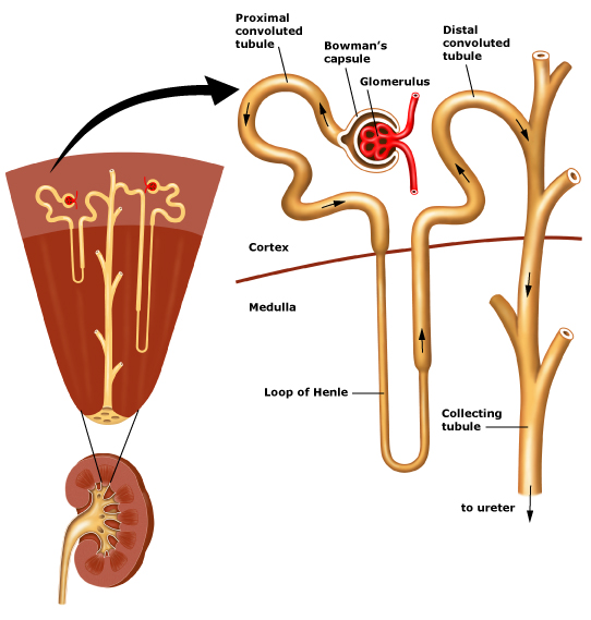
Position of the nephron in the kidney
|
Each kidney is made up of microscopic, coiled tube like units called nephrons or uriniferous tubules.
There are approximately 1.25 to 1.5 million nephrons in one kidney. The nephron has two parts-
The cortex has a dotted appearance because of the presence of these Malpighian corpuscle. The striated appearance of the medulla is due to the renal tubule. Nephrons that are present in the cortex region are called cortical nephrons, and the ones that extend to the medulla are called juxta medullary nephrons. |
http://www.biologycorner.com/anatomy/urinary/nephron01.jpg
|
labelling the diagram
|
http://www.edu.pe.ca/threeoaks/teacherpages/higginbotham/Biology%20521%20Webpage/resources/chapter12images/nephron4.jpg
|
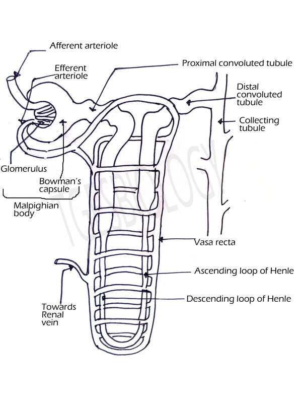
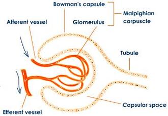
PARTS OF THE NEPHRON
Malpighian corpuscle has
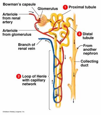
Each afferent arteriole leads to a network of capillaries called a glomerulus.
Blood leaves the capillaries of the glomerulus via an efferent arteriole and enters capillaries in the medulla called peritubular capillaries, which collect much of the water that was lost through the glomerulus.
Venules from the peritubular capillaries lead to the renal vein, which exits the kidney and returns blood to the inferior vena cava.
- A cup shaped - Bowman's capsule
- A network of capillaries- glomerulus
Each afferent arteriole leads to a network of capillaries called a glomerulus.
Blood leaves the capillaries of the glomerulus via an efferent arteriole and enters capillaries in the medulla called peritubular capillaries, which collect much of the water that was lost through the glomerulus.
Venules from the peritubular capillaries lead to the renal vein, which exits the kidney and returns blood to the inferior vena cava.
Drawing the Nephron
|
|
http://images.tutorvista.com/content/excretion/malpighian-corpuscle--structure.jpeg
|
|
RENAL TUBULE
The tubular part of the nephron is called as renal tubule. It has three parts- Proximal convoluted tubule, Loop of Henle and distal convoluted tubule. The proximal part of the renal tubule is highly coiled and is present in the renal cortex. This part absorbs water and all the useful salts, amino acids and glucose. The proximal part extends downwards to form a narrow tubular part- the descending part, a loop and an ascending part. This lies in the medulla and mainly absorbs water from the nephric filtrate. The ascending limb leads to a convoluted part the which also lies in the cortex. This opens into the collecting tubule. This part excretes some ions and ammonia into the filtrate. |
http://www.uaz.edu.mx/histo/pathology/ed/ch_16/c16_snephron.gif
|
|
URETER
URINARY BLADDER
URETHRA
|
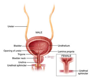
http://www.urologyhealth.org/urology/articles/images/anatomy_UretersBladder_coronal.jpg
|
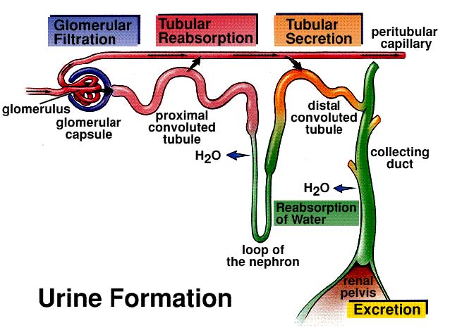
URINE FORMATION
|
ULTRAFILTRATION, RE-ABSORPTION AND TUBULAR SECRETION
URINE FORMATION TAKES PLACE IN THE KIDNEY. INVOLVES THREE PROCESSES:
|
http://fau.pearlashes.com/anatomy/Chapter%2041B/images/Chapter%2041B_img_1.jpg
|
Functions of the kidney-
- Excretion of nitrogenous wastes.
- Removal of excess of water and salts.
- Osmoregulation. (Osmoregulation is the control of the levels of water and mineral salts in the blood.)
URINE EXCRETION
Urine is formed by the combined action of glomerular filtration, tubular re-absorption and tubular secretion.
The urine is carried to the urinary bladder and when sufficient amount of urine is accumulated, urine is excreted out of the body.
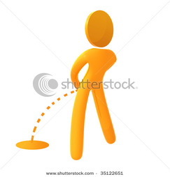
The act of voiding out of urine is called urination or micturation.
COMPOSITION OF URINE
Transparent pale yellow fluid acidic in nature.
Contains- water, mineral salts (NaCl), urea, uric acid, creatine and some excess hormones vitamins etc.
Smell is due to urinod, after a long time it smells of ammonia. Colour is due to urochrome
PATHOLOGICAL CONDITION
Urine is formed by the combined action of glomerular filtration, tubular re-absorption and tubular secretion.
The urine is carried to the urinary bladder and when sufficient amount of urine is accumulated, urine is excreted out of the body.
The act of voiding out of urine is called urination or micturation.
COMPOSITION OF URINE
Transparent pale yellow fluid acidic in nature.
Contains- water, mineral salts (NaCl), urea, uric acid, creatine and some excess hormones vitamins etc.
Smell is due to urinod, after a long time it smells of ammonia. Colour is due to urochrome
PATHOLOGICAL CONDITION
- Glycosauria- sugar in urine
- Ketonuria- ketone bodies in urine
- Pyuria- pus cells in urine
- Hematuria- blood in urine
|
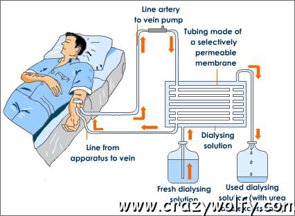
DIALYSIS
When the kidneys fail to function then the blood of the person is filtered through a dialysis machine or dialyser. It removes excess urea and salts from the blood such that the blood is filtered artificially. It is also called artificial kidney or kidney machine. |
|
