ISC 12>STRUCTURE AND FUNCTION OF PLANTS>1. PLANT ANATOMY> Plant tissues
Scope of syllabus
|
Class Presentation |
Plant body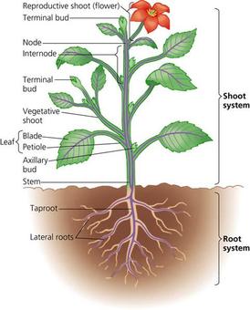
|
Plant tissues What do you mean by tissues?
A group of cells with similar origin, structure and function is called tissue. 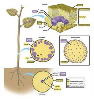
|
CLASSIFICATION OF PLANT TISSUES
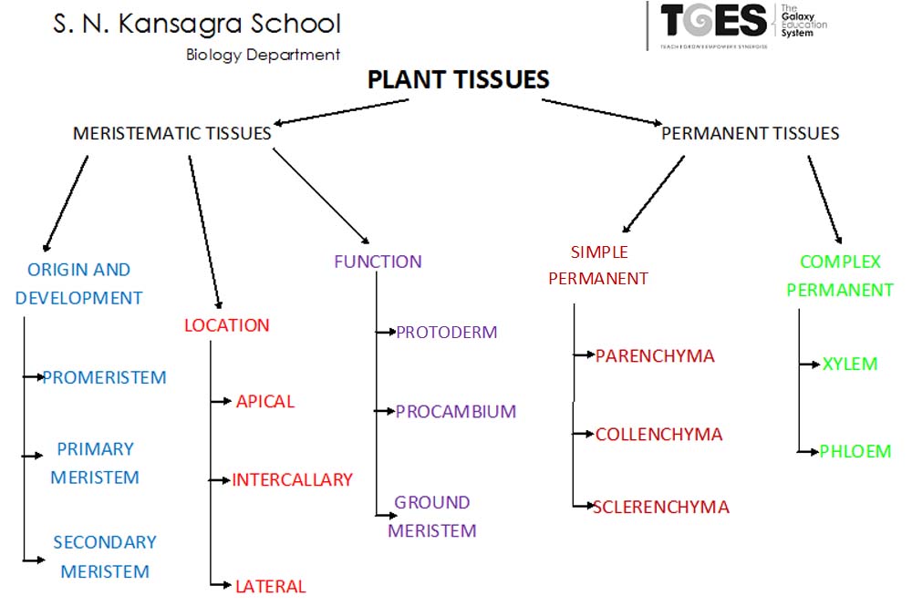
Meristematic tissue:
Define meristematic tissues.
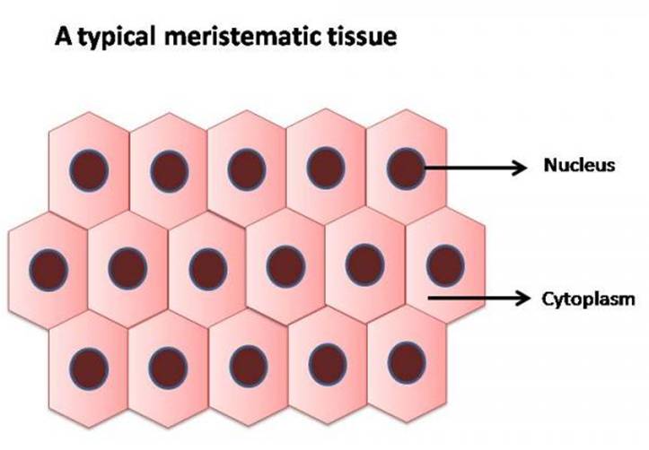
Meristematic tissue may be defined as a group of living cells which are located at specific location and divide continuously to add new cells to the plant body.
Meristematic tissue may be defined as a group of living cells which are located at specific location and divide continuously to add new cells to the plant body.
|
Characteristics of meristematic tissues.
|
|
Classification of meristematic tissue
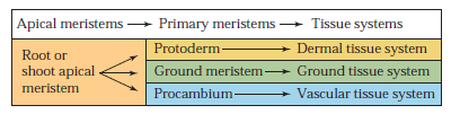
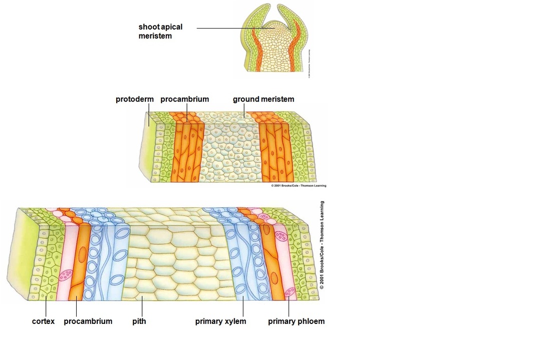
A) Based on Origin and development
(A) Promeristem:
(A) Promeristem:
- The meristem where foundation of new organs or of their part is laid down is called promeristem.
- They occupy very small area at the tips of stem and root.
- It differentiates into primary meristem.
- It originate from promeristem.
- Remains meristematic from the embryonic condition throughout entire plant life at the growing apices of roots, stems, primordial of leaves.
- Both apical meristems and intercalary meristems are primary meristems because they appear early in life of a plant and contribute to the formation of the primary plant body.
- It develops from primary permanent tissue.
- Fascicular vascular cambium, interfascicular cambium and cork-cambium.
- Responsible for producing the secondary tissues.
B)Based on location in plant body
|
1. Apical meristem
|
|
|
C)Based on function
|
|
|
|
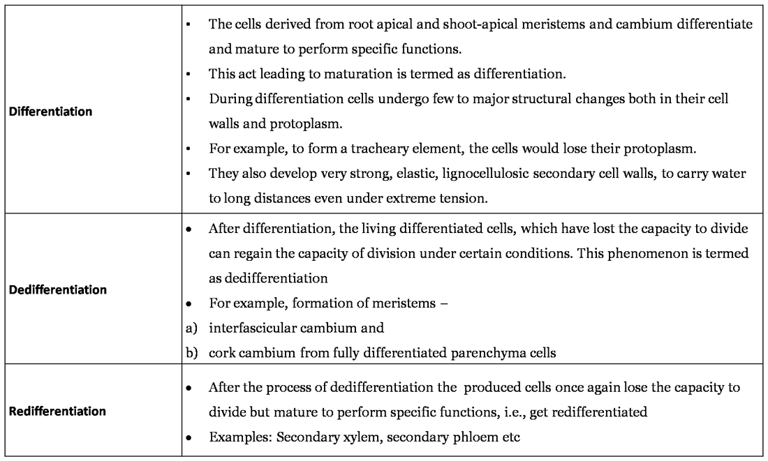
A BRIEF IDEA ABOUT DIFFERENTIATION, DEDIFFERENTIATION AND REDIFFERENTIATION
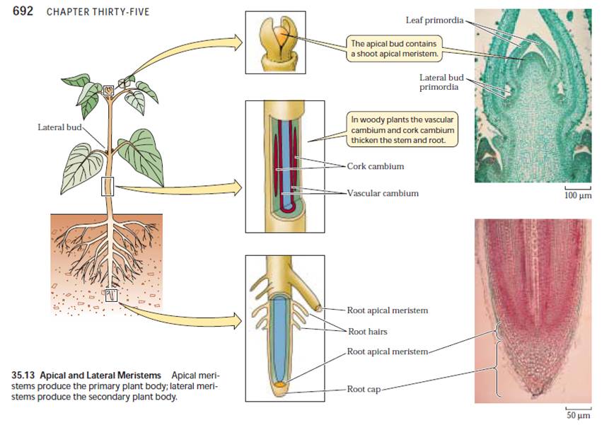
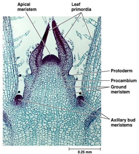
Shoot apex
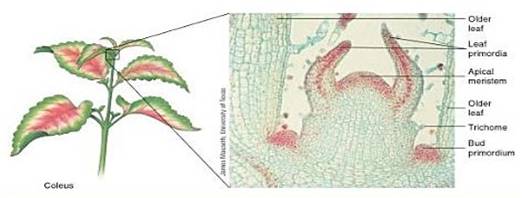
What do you mean by shoot apex....?
- Shoot apex is the portion of shoot above the youngest primordium.
- It is present at the plumular tip or at the end of leaf.
- It is covered by young leaves and visible on removing them.
- The apex is dome shaped.
Theories of shoot apex organisation:
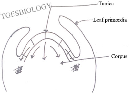
1. Tunica corpus theory :(Schmidt-1924)
1. Tunica corpus theory :(Schmidt-1924)
|
Observe the location of tunica and corpus regions

|
Draw the diagram
|
- Tunica corpus theory was given for vegetative shoot apex.
- According to this theory, there are two zones of tissues in the apical meristems.
- The tunica (Tunic = cover) consisting of one or more layers of peripheral layers of cells, and the corpus (corpus = body) a mass of cells enclosed by the tunica.
- The layers of tunica show anticlinal (perpendicular to periphery) divisions and bring about surface growth.
- In the corpus, cell division is irregular and at various planes resulting in growth and volume of the mass.
- Tunica gives rise to epidermis.
- Corpus gives rise to Cortex/ground tissue.
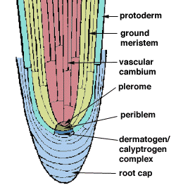
2. Histogen Theory (Hanstein 1870)
(ii) Periblem: (Middle layer) Gives rise to cortex and endodermis.
(iii) Plerome: (Inner layer): gives rise to pericycle, pith and vascular tissue.
Shoot apex: Labeling exercise
- According to this theory, shoot apex has three zones.
- Apical meristem of stem composed of small mass of cells which are all alike and divide fast (meristematic).
- These meristematic cells form promeristem, which differentiate into three zones : dermatogen, periblem and plerome.
- Every zone consists of a group of initials called a histogen (tissue builder).
(ii) Periblem: (Middle layer) Gives rise to cortex and endodermis.
(iii) Plerome: (Inner layer): gives rise to pericycle, pith and vascular tissue.
Shoot apex: Labeling exercise
Root apex
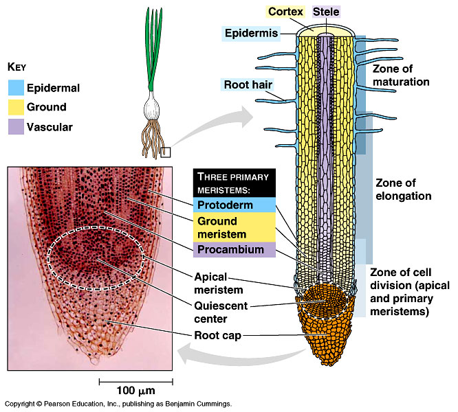
What do you mean by root apex....?
- Root apex has simplest organisation as compared to stem.
- Root apex is sub terminal in position because of terminal position of root cap.
|
Theories of root apex organisation:
1. Histogen Theory (Hanstein 1870)
(ii) Periblem: (Middle layer) Gives rise to cortex and endodermis. (iii) Plerome: (Inner layer): gives rise to pericycle, pith and vascular tissue. Calyptrogen:
|
|
Quiescent centre
- Quiescent centre concept given by Clowes (1956) in maize.
- According to this, there is an inactive centre in the root apex which is called quiescent centre (having low DNA, RNA, protein) and it acts as a reservoir of active initials.
Calyptra and calyptrogen
- Calyptra: Root cap which protects the root tip.
- Calyptrogen: Meristematic tissue which produce calyptra/root cap.
Permanent tissues
Permanent tissue may be defined as a group of living or dead cells formed by meristematic tissue and have lost their ability to divide and have permanently placed at fixed position in plant body.
I) Simple permanent tissues
II) Complex permanent tissues
I) Simple permanent tissues
II) Complex permanent tissues
I) Simple permanent tissues
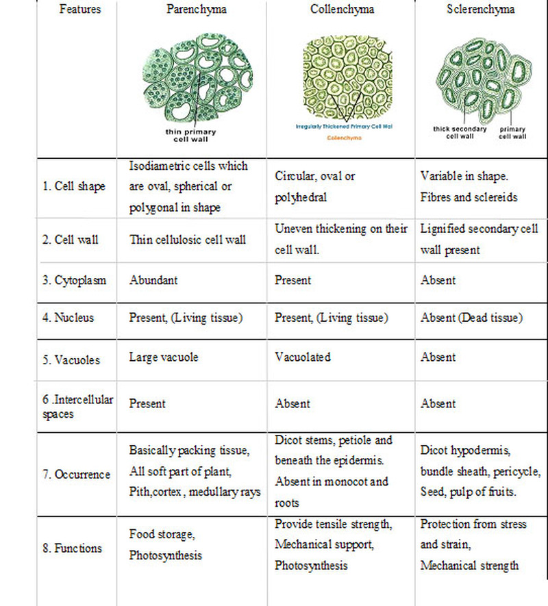
TYPES OF SCLERENCHYMA
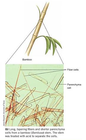
i) Fibres
|
ii) Sclereids ( Also known as stone cells or sclerotic cells) Occurrence : Most common in fruits and seeds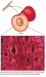
A) Structure
. |
II)Complex permanent tissue
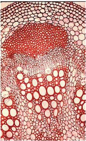
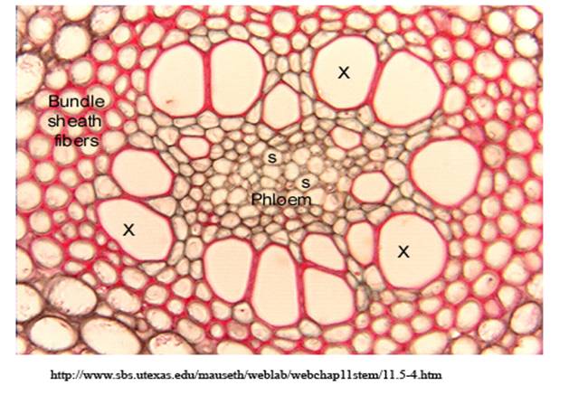
A) Xylem :
i) Distribution in plant body:
Xylem & phloem in root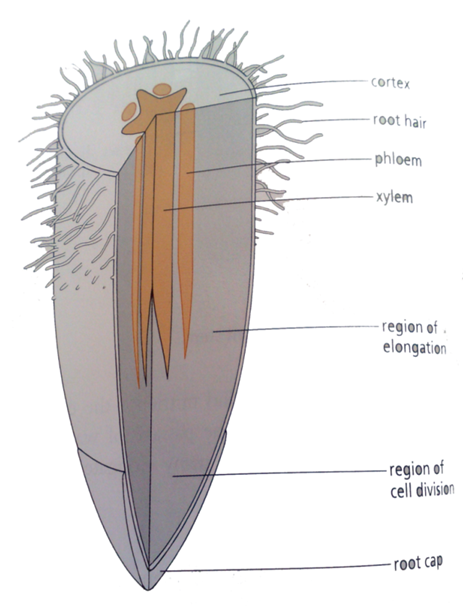
|
Xylem & phloem in stem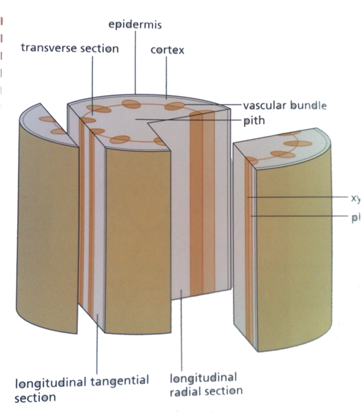
|
Xylem & phloem in leaf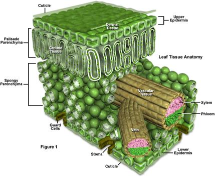
|
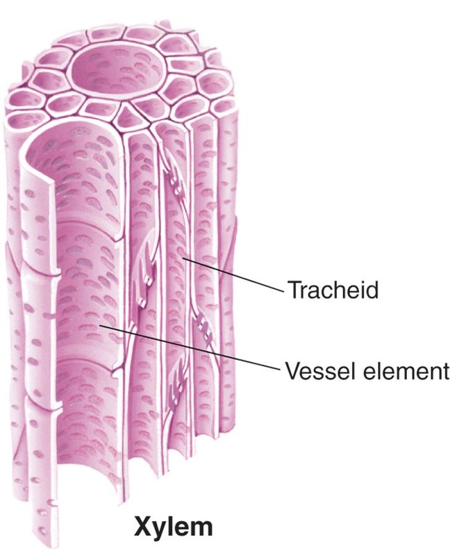
ii) Components of xylem:
|
Components:
|

|
|
Protoxylem and metaxylem
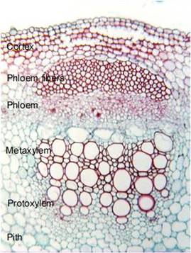
|
Difference between protoxylem and metaxylem

|
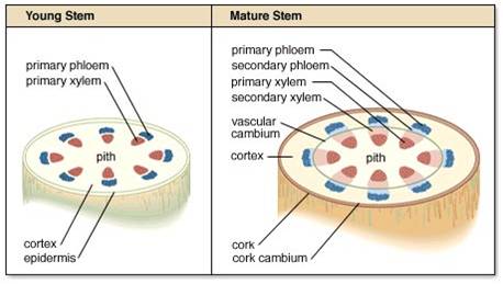
- Primary and secondary xylem

|
|
Types of xylem : Endarch, exarch and mesarch
Based on relative position of protoxylem and metaxylem, the xylem is of the following three types.
Based on relative position of protoxylem and metaxylem, the xylem is of the following three types.
- Exarch: A condition in which the protoxylem lies towards the periphery and metaxylem towards the central axis. (Root)
- Endarch: A condition in which protoxylem lies towards the centre and metaxylem towards the periphery.(Stem)
- Mesarch: A condition in which metaxylem remain in the centre surrounded all around by the protoxylem.
B. Phloem
i) Distribution in plant body: (observe the figures given in xylem distriution)
i) Distribution in plant body: (observe the figures given in xylem distriution)
ii) Components of phloem :
|
Components:
|
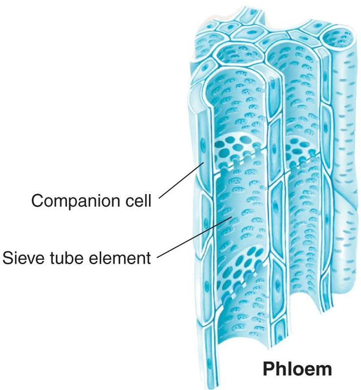
|
Sieve cell
|
Primary phloem and secondary phloem

|

|
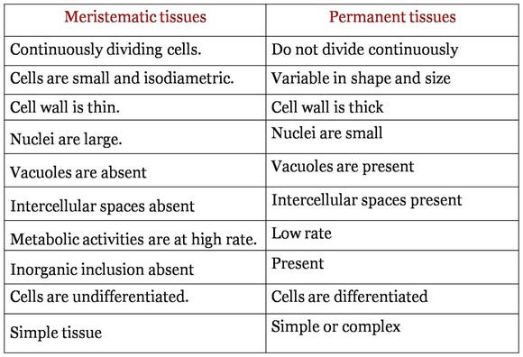
Differences between meristematic and permanent tissues
Types of vascular bundle
A. Based on cambium
2 Types
- Open vascular bundle: Vascular bundle with cambium.
- Closed vascular bundle : Vascular bundle without cambium.
DRAW THE DIAGRAM
B. Based on xylem and phloem arrangement
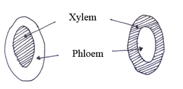
1. Radial vascular bundle:
|
In type of arrangement xylem and phloem form separate bundles alternating with each other i.e. they lie on different radii.
This arrangement is the characteristic of root. 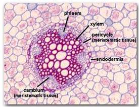
|
DRAW THE DIAGRAM
Radial vascular bundle 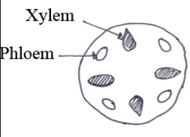
|
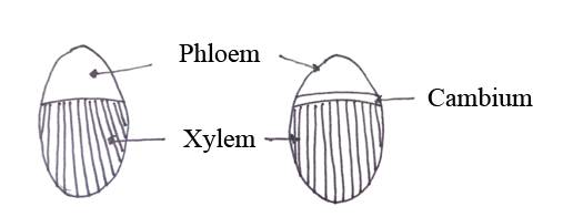
2. Conjoint vascular bundle
Conjoint: When xylem and phloem are combine into one bundle.
a) Collateral: When xylem and phloem are situated at the same radius of vascular bundles.
b) Bicollateral: In this phloem and cambium lie on both sides of xylem. (Eg. Cucurbitaceae )
a) Collateral: When xylem and phloem are situated at the same radius of vascular bundles.
b) Bicollateral: In this phloem and cambium lie on both sides of xylem. (Eg. Cucurbitaceae )
|
A) Conjoint collateral
|
DRAW THE DIAGRAM
Conjoint collateral 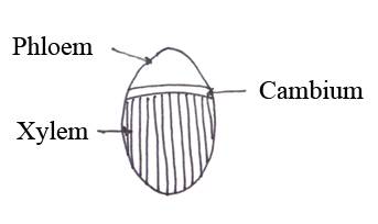
|
b) Conjoint bicollateral

|
DRAW THE DIAGRAM
Conjoint bicollateral 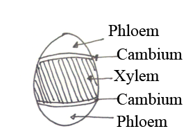
|
3.Concentric vascular bundle
When xylem and phloem arrange concentrically, i.e. one surrounds the other completely.
a) Amphicribal: When xylem lies in the centre and is completely surrounded by the phloem. (Ferns)
b) Amphivasal: When phloem lies in centre and is completely surrounded by the xylem. (Some monocot stem)
a) Amphicribal: When xylem lies in the centre and is completely surrounded by the phloem. (Ferns)
b) Amphivasal: When phloem lies in centre and is completely surrounded by the xylem. (Some monocot stem)
|
A) Amphicribal
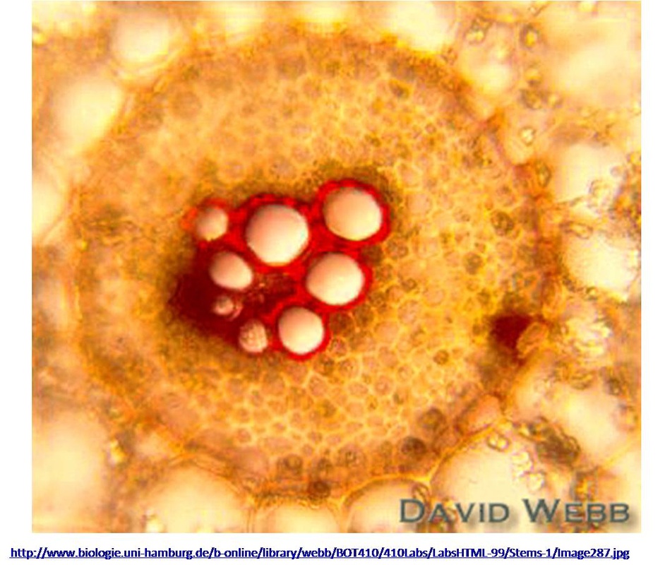
|
DRAW THE DIAGRAM
Amphicribal & amphivasal vascular bundles
|
B) Amphivasal
|
Anatomical differences between dicot and monocot root, stem and leaf
ASSESSMENT
|
| ||||||||
