ISC 12> UNIT 2> STRUCTURE AND FUNCTION OF ANIMALS> HUMAN REPRODUCTION
SCOPE OF SYLLABUS
HUMAN REPRODUCTION-
Organs of male and female reproductive system and their functions;
internal structure of testis and ovary to be taught with the help of diagrams;
gametogenesis- spermatogenesis (including spermiogenesis and spermiation)
oogenesis; hormonal control of gametogenesis,
structure of sperm and mature ovum,
menstrual cycle - different phases and hormone action, differences between oestrous and menstrual cycle, menarche and menopause.
CHANGED SYLLABUS [2020-21]
Internal structure of testis and ovary to be taught with the help of diagrams;
gametogenesis- spermatogenesis (including spermiogenesis and spermiation)
oogenesis; hormonal control of gametogenesis,
structure of sperm and mature ovum,
menstrual cycle - different phases and hormone action, menarche and menopause,
gametogenesis- spermatogenesis (including spermiogenesis and spermiation)
oogenesis; hormonal control of gametogenesis,
structure of sperm and mature ovum,
menstrual cycle - different phases and hormone action, menarche and menopause,
|
Reproduction is defined as a biological process in which an organism gives rise to young ones (offspring) similar to itself.
The offspring grow, mature and in turn produce new offspring. Thus, there is a cycle of birth, growth and death. Reproduction enables the continuity of the species, generation after generation. Based on whether there is participation of one organism or two in the process of reproduction, it is of two types. When offspring is produced by a single parent with or without the involvement of gamete formation, the reproduction is asexual. When two parents (opposite sex) participate in the reproductive process and also involve fusion of male and female gametes, it is called sexual reproduction The primary sex organs are called gonads. Primary organs are testes in male and ovaries in females. The testis produces sperms and the male hormone testosterone The ovaries produces ova and the female hormone estrogen and progesterone |
|
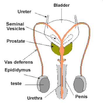
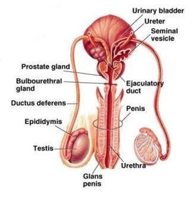
MALE REPRODUCTIVE SYSTEM
Parts of the male reproductive system
|
The male reproductive system is located in the pelvis region. It includes a pair of testes along with accessory ducts, glands and the external genitalia.
The primary reproductive organ- The testes are situated outside the abdominal cavity within a pouch called scrotum. The testes descend through the inguinal canal into the scrotum at the time of birth. FUNCTION: The scrotum helps in maintaining temperature of the testes 2–2.5 degree C lower than the normal internal body temperature. This is necessary for spermatogenesis. The failure of testes to descend into the scrotal sac is called chryptorchidism The internal organs of the male reproductive system, also called accessory organs, include the following: The epididymis is a long, coiled tube that rests on the backside of each testicle. It transports and stores sperm cells that are produced in the testes. It also brings the sperm to maturity, since the sperm that emerge from the testes are immature and incapable of fertilization. The vas deferens is a long, muscular tube that travels from the epididymis into the pelvic cavity, to just behind the bladder. The vas deferens transports mature sperm to the urethra, the tube that carries urine or sperm to outside of the body, in preparation for ejaculation. Ejaculatory ducts are formed by the fusion of the vas deferens and the seminal vesicles (see below). The ejaculatory ducts empty into the urethra. Urethra is the tube that carries urine from the bladder to outside of the body. In males, it has the additional function of ejaculating semen when the man reaches orgasm. When the penis is erect during sex, the flow of urine is blocked from the urethra, allowing only semen to be ejaculated at orgasm. |
THE ACCESSORY GLANDS
|
|
Structure of testes
|
In adults, each testis is oval in shape, with a length of about 4 to 5 cm and a width of about 2 to 3 cm. The testis is covered by a dense covering.
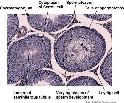
The tunica vaginalis is the outer serous covering of the testis. The Tunica Albuginea is the inner fibrous covering of the testis. It is a dense membrane, of a bluish-white color, composed of bundles of white fibrous tissue which interlace in every direction. It is covered by the tunica vaginalis, except at the points of attachment of the epididymis to the testis, and along its posterior border, where the spermatic vessels enter the gland.From the inners surface of the tunica albuginea, a series of fibrous septa extend towards the interior of the organ. These septa divide the testicle into several lobules. Each testis has about 250 compartments called testicular lobules. Within each lobule, there lay one to three coiled tubules known as seminiferous tubules. The regions outside the seminiferous tubules called interstitial spaces, contain small blood vessels and interstitial cells or Leydig cells. Leydig cells synthesise and secrete testicular hormones called androgens. The seminiferous tubules open into a network of channels called the rete testis. Small efferent ductules connect the rete testis to the upper end of the epididymis. |
|
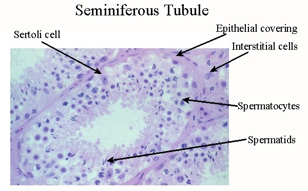
Seminiferous tubule
|
|
|
|
Each seminiferous tubule consists of a basement layer formed of laminated connective tissue containing numerous elastic fibers with flattened cells between the layers and covered externally by a layer of flattened epithelioid cells.
Within the basement membrane are epithelial cells arranged in several irregular layers. Among these cells may be seen the spermatozoa in different stages of development. (1) Lining the basement membrane and forming the outer zone is a layer of cubical cells, with small nuclei; some of these enlarge to become spermatogonia. (2) Within this next layer is a number of larger polyhedral cells, with clear nuclei, arranged in two or three layers; these are the intermediate cells or spermatocytes. Most of these cells are in a condition of karyokinetic division, and the cells which result from this division form those of the next layer, the spermatoblasts or spermatids. (3) The third layer of cells consists of the spermatoblasts or spermatids, and each of these, without further subdivision, becomes a spermatozoon. The spermatids are small polyhedral cells, the nucleus of each of which contains half the usual number of chromosomes. In addition to these three layers of cells others are seen, which are termed the supporting cells (cells of Sertoli). They are elongated and columnar, and project inward from the basement membrane toward the lumen of the tube. As development of the spermatozoa proceeds the latter group themselves around the inner extremities of the supporting cells. |
|
External genitilia- Penis
The penis is the male external genitalia, made up of special tissue that helps in erection of the penis to facilitate insemination. The enlarged end of penis called the glans penis is covered by a loose fold of skin called foreskin.
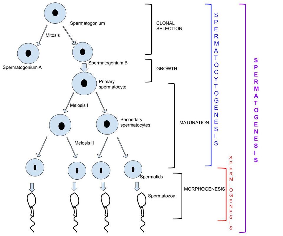
Spermatogenesis
Spermatogenesis
|
The development of the germ cells begins with the spermatogonia at the periphery of the seminal canal and advances towards the lumen.
The cells form spermatocytes I (primary spermatocytes), spermatocytes II (secondary spermatocytes), spermatids and finally to mature sperm cells. Multiplication phase (spermatocytogenesis): The germinal epithelium divides mitotically to produce sperm mother cells called spermatogonium. In this division two daughter cells are formed - one remains as spermatogonium and the other proceeds to form the primary spermatocyte. The primary spermatocyte undergoes meiosis. |
|
Process of spermatogenesis
|
Growth phase: The spermatocyte and the nucleus enlarges and gets ready for the maturation phase.
Maturation phase: The primary spermatocytes undergo meiosis to form secondary spermatocytes with half the number of chromosomes. The secondary spermatocytes form spermatids.The spherical spermatid cells undergo differentiation to form the sperms. The differentiation of the spermatids to form sperms is called as spermiogenesis. The spermatozoa formed are fully developed but are not entirely motile. They derive nourishment from the sertoli cells and are released to the lumen. This process of release of sperms from the sertoli cells is called spermiation. These are pushed to the epididymis and attain full motility in the cauda epididymis. Significance: One spermatogonium produces four sperms. Meiosis reduces the chromosome number to half and thus maintains the chromosome number of the species. Meiosis also produces variations. |
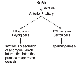
HORMONAL CONTROL OF SPERMATOGENESIS
|
Spermatogenesis starts at the age of puberty due to significant increase in the secretion of gonadotropins.
The increased levels of GnRH then acts at the anterior pituitary gland and stimulates secretion of two gonadotropins – follicle stimulating hormone and intestitial cell stimulating hormone. ICSH acts on the Leydig cells and stimulates synthesis and secretion of androgens. Androgens, in turn, stimulate the process of spermatogenesis. FSH acts on the Sertoli cells and stimulates secretion of some factors which help in the process of spermiogenesis. |
Sperm cell
|
|
A mature spermatozoan is approximately 60um long, flagellated, motile cell. It consists of flat oval head and narrow tail.
Head: It is flat and oval and consists of (1) anterior, acrosome that contains hydrolytic enzymes. These enzymes help in penetrating the egg membrane during fertilization and (2) posterior, large nucleus that contains a haploid set of chromosomes. Mid piece: It consists of a sheath of ring-shaped mitochondria grouped around the axoneme to provide the energy for the flagellar movement. Tail: The tail has 3 parts- 1. A narrow neck with two centrioles, 2. The middle piece which is composed of many mitochondria forming a spiral, that provides energy for to the sperm for motility. 3. The principal piece of the tail that finally ends in end piece. |
The ejaculate
The human male ejaculates about 200 to 300 million sperms during coitus of which, for normal fertility, at least 60 per cent sperms must have normal shape and size and at least 40 per cent of them must show vigorous motility.
The human male ejaculates about 200 to 300 million sperms during coitus of which, for normal fertility, at least 60 per cent sperms must have normal shape and size and at least 40 per cent of them must show vigorous motility.
FEMALE REPRODUCTIVE SYSTEM
|
|
|
|
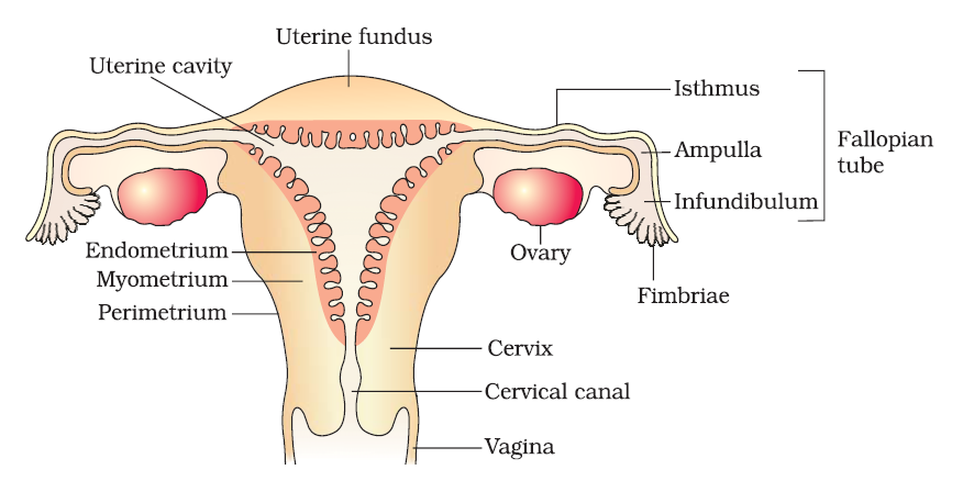
The female reproductive system consists of a pair of ovaries alongwith a pair of oviducts, uterus, cervix, vagina and the external genitalia located in pelvic region. These parts of the system alongwith a pair of the mammary glands are integrated structurally and functionally to support the processes of ovulation, fertilisation, pregnancy, birth and child care.
Ovary: Ovaries are the primary female sex organs that produce the female gamete (ovum) and several steroid hormones (ovarian hormones). They are located one on each side of the lower abdomen Each ovary is about 2 to 4 cm in length and is connected to the pelvic wall and uterus by ligaments. |
Oviduct/fallopian tube: The oviducts, uterus and vagina constitute the female accessory ducts. Each fallopian tube is about 10-12 cm long and extends from the periphery of each ovary to the uterus, the part closer to the ovary is the funnel-shaped infundibulum. The infundibulum possess finger-like projections called fimbriae, which help in collection of the ovum after ovulation. The infundibulum leads to a wider part of the oviduct called ampulla. The last part of the oviduct, isthmus has a narrow lumen and it joins the uterus.
Uterus: The uterus is single and it is also called womb. It is supported by ligaments attached to the pelvic wall. The uterus opens into vagina through a narrow cervix. The cavity of the cervix is called cervical canal which along with vagina forms the birth canal. The wall of the uterus has three layers of tissue. The external thin membranous perimetrium, middle thick layer of smooth muscle, myometrium and inner glandular layer called endometrium that lines the uterine cavity.
Vagina: Vagina is a strong muscular tube that connects the cervix of the uterus to the vestibule outside. It is about 3 to 5 inches long. The cavity of the cervix, the cervical canal and the vagina forms the birth canal. Vagina also receives the sperms during coitus.
|
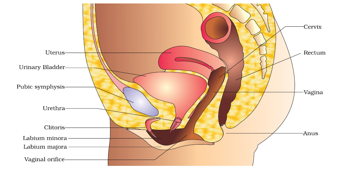
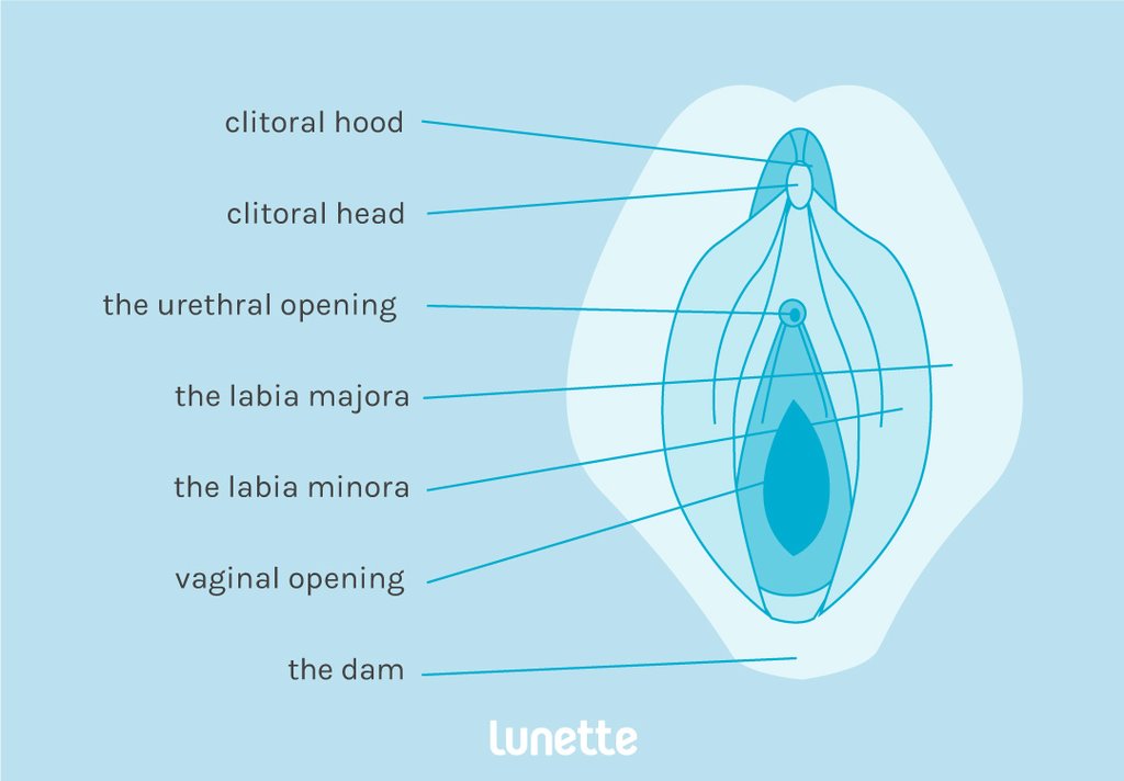
Vulva- The external genitalia-
The mons pubis, a fatty tissue with pubic hair. The folds of muscular tissue around the vaginal opening include two thick folds labia majora and two small folds labia minora these protects the inner parts. A small protrusion of tissue covered with a thin flap of tissue is clitoris, which is the main source of sensations during sexual activity. Vestibule is a depression that forms a small cavity of the vulva, that has two openings –
Bartholin’s gland These are two bean shaped gland, on either side of the vaginal orifice. They secrete lubricating fluid. |
|
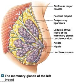
The mammary gland
|
The mammary glands are paired structures (breasts) that contain glandular tissue and variable amount of fat. These are modified sweat glands.
It is in the form of protuberance on the chest and are called breasts. These glands begin to develop at puberty, under the effect of gonadotropins. The glandular tissue of each breast is divided into 15-20 mammary lobes containing clusters of cells called alveoli. The cells of alveoli secrete milk, which is stored in the cavities (lumens) of alveoli. The alveoli open into mammary tubules. The tubules of each lobe join to form a lactiferous ducts. Several mammary ducts join to form a wider mammary ampulla which is connected to lactiferous duct through which milk is sucked out. Each breast has and erectile nipple, surrounded by a dark pigmented area called the areola. There are sebaceous glands called areolar glands in the areola. |
|
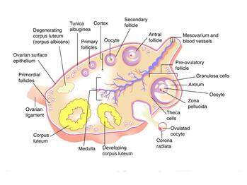
INTERNAL STRUCTURE OF OVARY
The surface of the ovary is covered by a layer of columnar cells which constitutes the germinal epithelium. There is a layer of connective tissue on the outer surface called the tunica albuginea.
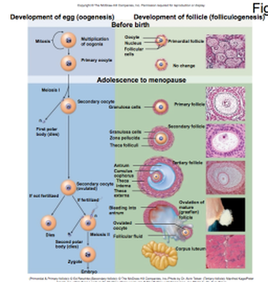
Internally the ovary consists of a number of vesicular ovarian follicles embedded in the meshes of a stroma. The stroma of the ovary may contain interstitial cells resembling those of the testis, and is supplied with abundant blood vessels. The developing follicles are present near the periphery called as cortical stroma and the highly vascularised inner region called medullary stroma. The follicles are spherical aggregations of cells in the ovary that contains a single oocyte. These follicles grow from a small primordial follicle to a mature Graafian follicle. This process is called folliculogenesis.
Internally the ovary consists of a number of vesicular ovarian follicles embedded in the meshes of a stroma. The stroma of the ovary may contain interstitial cells resembling those of the testis, and is supplied with abundant blood vessels. The developing follicles are present near the periphery called as cortical stroma and the highly vascularised inner region called medullary stroma. The follicles are spherical aggregations of cells in the ovary that contains a single oocyte. These follicles grow from a small primordial follicle to a mature Graafian follicle. This process is called folliculogenesis.
|
|
|
|
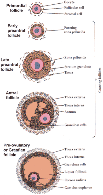
Basic types of ovarian follicle include:
Primordial follicle: These appear in the third month of foetal development. The oocyte is surrounded by a single layer of squamous follicular cells. Primary follicle- has a central oocyte surrounded by a single layer of cuboidal cells. These cells are called granulosa cells. The zona pellucida is also visible. Secondary follicle- The characteristic feature of this is development of cavities in-between the granulosa cells. Outer layer of theca is visible. Graffian follicle- This stage is characterized by the presence of a large follicular cavity or the antrum filled with liquor folliculi. The thecal layers become more prominent and the layer of granulosa cells become thinner. Atretic follicle: Some of the developing follicles degenerate during the process of folliculogenesis. Corpus haemorrhagicum: a ruptured graafian follicle containing a blood clot that is absorbed as the cells lining the follicle form the corpus luteum. Corpus luteum: The corpus haemorrhagicum, formed after rupturing of the follicle changes to form a yellow coloured mass of cells called corpus luteum. This structure secretes the hormone progesterone during the luteal phase. Corpus albicans: When the oocyte is not fertilised, the corpus luteum starts to degenerate and stops secreting progesterone. This then forms a mass of fibrous scar tissue and is ca |
STRUCTURE OF GRAAFIAN FOLLICLE
|
|
Graafian follicle is a mature ovarian follicle. This characterised by the presence of a large cavity called as antrum. The antrum is filled with a fluid called liquor folliculi.
The oocyte increases in size and is surrounded by a row of cells called the corona radiata. The oocyte has undergone the first meiotic division and is arrested in the metaphase stage. The oocyte shifts to a side and lies on a mass of cells called cumulus oophorous. The granulosa cells form a thin layer of cells towards the periphery. The connective tissue layers outside the follicle differentiate to form two distinct layers- the theca interna and the theca externa. |
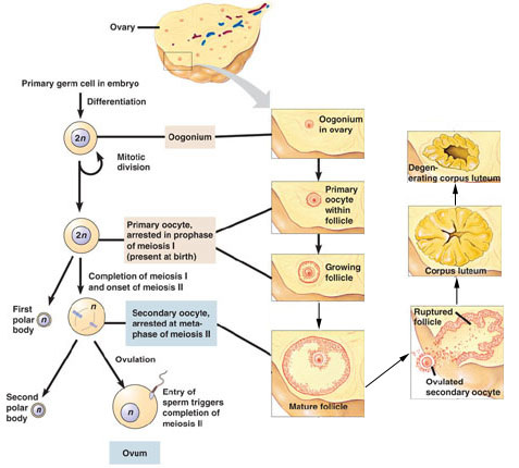
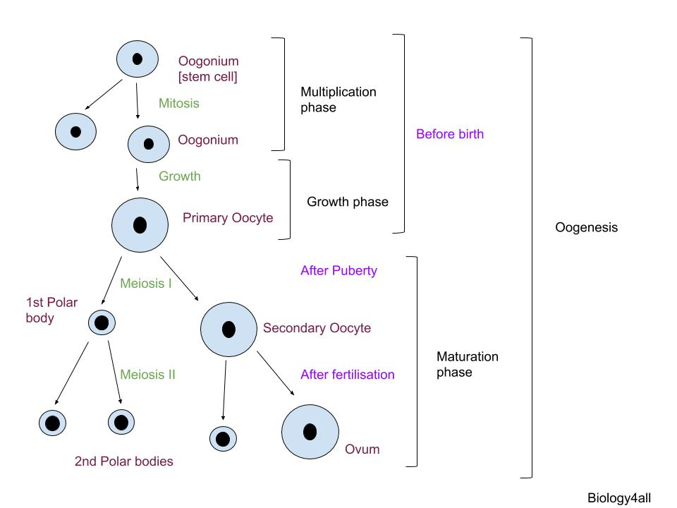
OOGENESIS
|
The process of egg formation or oogenesis begins before the birth of the female.
Multiplication phase: The germinal epithelium produces many oogonia that develop to form the primary oocytes. These are cells that become larger and has large nucleus. These cells form a mass called egg nest. One cell enters the growth phase whereas other cells form the follicle cells surrounding the oocyte. Growth phase: The primary oocyte divides by meiosis to form two unequal cells- (1) cell with large amount of cytoplasm is the secondary oocyte and (2) cell with very less cytoplasm called the polar body. This includes vitellogenesis, formation of zone pellucida and increased activity of different organelles like mitochondria, golgi body etc. There will be around 2 million primary oocytes in the ovary of a new born female. Maturation phase: The secondary oocyte undergoes meiosis II and is arrested in the metaphase stage till ovulation. The division is complete only if fertilisation takes place. This gives rise to the ovum and another polar body. The polar bodies disintegrate. |
Oogenesis- the process
Oogenesis in human female begins during the embryonic phase at 5 months of gestation. There is a peak of about 6-7 million oogonia, after 5-6 months production of oogonia stops and never resumes. The specialised cells that undergo meiosis to form ovum are oogonium .
These oogonia become primary oocyte by the end of gestation, (birth of the female)
At puberty there are only about 400,000 primary oocytes. The process of oogenesis continues at puberty due to the pituitary gonadotropin.
Oogenesis stops at the age of 45-50. Only about 400 of these oocytes will actually ovulate during a woman's lifetime.
The oocyte is present within the follicle, and as the follicle develops oogenesis takes place.
Primary oocyte arrested in meiosis I is present in primary, secondary follicle and tertiary follicle.
The primary oocyte within the tertiary follicle grows in size and completes its first meiotic division.
It is an unequal division resulting in the formation of a large haploid secondary oocyte and a tiny first polar body. It remains arrested in meiosis II as secondary oocyte when the follicle ruptures, and completes meiosis II only after fertilisation.
Oogenesis - Diagram |
How to draw |
|
|
SPERMATOGENESIS1. Process takes place in the seminiferous tubules.
2. Process begins at puberty. 3. Process continuous throughout the life time. 4. Four functional, motile sperms are formed after the process. 5. No polar bodies are formed during this process. 6. The spermatozoa produced are minute, stramlined and yolkless. |
OOGENESIS1. Process takes place in the ovaries.
2. Process begins in the fetus. 3. Process stops after menopause. 4. One functional, non-motile ovum is produced after the process. 5. Two to three polar bodies are produced during this process. 6. The ovum is large, round with yolk. |
FOLLICULOGENESIS AND OVULATION
|
The germinal epithelium divides to form follicle cells that surround a primary oocyte to form the primary follicle. At birth the baby girl will have about a million primary follicles. The primary oocyte starts the meiotic division but is arrested at prophase I. At puberty, hormones from the pituitary stimulate the follicle to develop further. Each month several follicles start to develop but usually only one matures to form a graafian follicle. The remaining undergo atresia. The follicle cells of the primary follicle multiply and a number of fluid filled spaces appear between them. This is now a secondary follicle. The secondary follicle forms a mature follicle and migrates to the surface of the ovary. Eventually the follicle bursts and the secondary oocyte with its surrounding cells is released in the out of the ovary. This process is called ovulation. After ovulation the remaining follicle cells develop to form the corpus luteum. |
STRUCTURE OF THE OVUM /OOTID
|
|
The ovum is the largest cell in the human body with a diameter of 14mm. It is spherical and non-motile. The cytoplasm has small lipid droplets and the cytoplasm is called as ooplasm. It has a nucleus called the germinal vesicle. The ovum is enclosed by a thick, transparent envelope called the zona striata or zona pellucida. This is a layer or mucopolysaccharide. The space between the plasma membrane and the zona pellucida is called peri-vitelline space. To the outer surface of the zona pellucida there are two to three strata of cells derived from the follicle called corona radiata. The human ovum does not contain yolk hence is called alecithal and is called non-cleidoic as it does not contain shell. |
Hormonal control of oogenesis
Gonadotropins regulate the process of folliculogenesis and oogenesis.
Follicle stimulating hormone stimulates the meiotic divisions and the granulosa cells to produce estrogen.
Luteinizing hormone stimulates ovulation and promotes the development of corpus luteum.
Follicle stimulating hormone stimulates the meiotic divisions and the granulosa cells to produce estrogen.
Luteinizing hormone stimulates ovulation and promotes the development of corpus luteum.
Significance of oogenesis
- Produces haploid ovum
- Most cytoplasm is retained in the functional cell
- Variations occur during meiotic division.
MENSTRUAL CYCLE
|
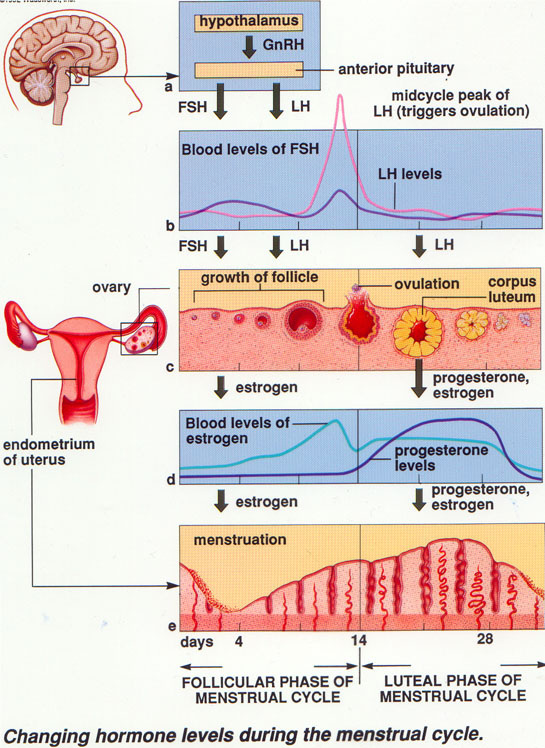
The reproductive cycle in the female primates (e.g. monkeys, apes and human beings) is called menstrual cycle.
The first menstruation begins at puberty and is called menarche. In human females, menstruation is repeated at an average interval of about 28/29 days, and the cycle of events starting from one menstruation till the next one is called the menstrual cycle. One ovum is released (ovulation) during the middle of each menstrual cycle. In human beings, menstrual cycles ceases around 50 years of age; that is termed as menopause. Cyclic menstruation is an indicator of normal reproductive phase and extends between menarche and menopause. CHANGES IN UTERUS Inner lining of the uterus, thickens or proliferates as the follicle develops in the ovary due to the action of estrogen hormone. The thickening is in anticipation of the pregnancy.If there is no fertilisation the thickened endometrium sheds off as menstrual fluid This process is called menstruation. |
|
Phases of the menstrual cycle
|
Menstrual phase (1st to 5th day)- In the absence of fertilisation, the corpus luteum degenerates. This causes disintegration of the endometrium leading to discharge of blood carrying broken uterine tissue. This flow of blood is called menstruation.
Follicular phase (Post menstrual or Proliferative phase) 6th to 13/14th day-During this phase, the primary follicles in the ovary grow to become a fully mature Graafian follicle and simultaneously the endometrium of uterus regenerates through proliferation. Endometrial glands elongate and become corkscrew shaped. The arterioles become longer and coiled. Epithelial cells of fallopian tube get thickened and movement of cilia increases. These changes in the ovary and the uterus are induced by changes in the levels of pituitary and ovarian hormones. The secretion of gonadotropins (LH and FSH) increases gradually during the follicular phase, and stimulates follicular development as well as secretion of estrogens by the growing follicles. Both LH and FSH attain a peak level in the middle of cycle (about 14th day). Ovulatory phase (13/14th day) -Rapid secretion of LH leading to its maximum level during the mid-cycle called LH surge induces rupture of Graafian follicle and thereby the release of ovum (ovulation). Luteal phase (Post ovulatory, secretory or premenstrual phase) 15th to 28th day- The ovulation (ovulatory phase) is followed by the luteal phase during which the remaining parts of the Graafian follicle transform as the corpus luteum. The corpus luteum secretes large amounts of progesterone which is essential for maintenance of the endometrium. Uterine glands enlarge and releases nutritional fluids. Such an endometrium is necessary for implantation of the fertilised ovum and other events of pregnancy. During pregnanacy all events of the menstrual cycle stop and there is no menstruation. |
|
What happens during the menstrual cycle?
1. The start of the menstrual cycle begins on the first day of bleeding. On about the 5th day, the brain begins producing hormones that cause a few follicles to grow and mature in the ovaries. These follicles then start producing the hormone estrogen.
2. The lining of the uterus (endometrium) responds to the estrogen by becoming thicker and developing more blood vessels.
3. Somewhere around the 14th or 15th day of the cycle, the most mature follicle bursts and releases an egg or ovum. This is called ovulation.
4. The egg then enters the Fallopian tube.
5. The ruptured follicle becomes the corpus luteum and begins to secrete the hormone progesterone.
6. Progesterone increases the supply of blood and nutrients to the uterus and maintains the endometrial lining.
7. If the egg unites with a sperm from a man, it becomes fertilized and travels down the Fallopian tube, finally settling into the lining of the uterus. If the egg does not become fertilized, it soon breaks down, and the follicle stops producing progesterone.
8. The loss of progesterone causes the lining of the uterus to break apart and bleed. The menstrual period then begins, starting another cycle.
Hormones and the menstrual cycle
|
|
