ISC 12> STRUCTURE AND FUNCTION OF ANIMALS> 8. LOCOMOTION
SCOPE OF SYLLABUS
Locomotion:
Joints, muscle movements, types of skeletal movements, basic aspects of human skeleton.
Functions of human skeleton;
different types of joints - their location and function;
Joints, muscle movements, types of skeletal movements, basic aspects of human skeleton.
Functions of human skeleton;
different types of joints - their location and function;
SKELETAL TISSUE (part of Chapter 1- Animal tissue)
|
It forms a strong frame work, that supports the body, protects the internal organs and provides surface for insertion of muscles.
This tissue occurs in two forms-
|
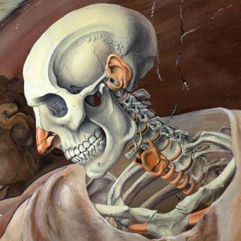
"Illustration by Michelle Annette Leveille" |
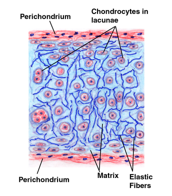
1. CARTILAGE
|
It has matrix made up of a protein chondrin or chondromucoid.
This is secreted by cartilage cells called chondrocytes. The chondrocytes are present in fluid filled spaces called lacunae. The tissue is surrounded by a membrane called perichondrium. Cartilage is classified into three types-
|
|
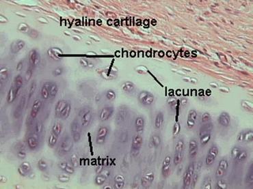
HYALINE CARTILAGE
|
Hyaline cartilage is glassy bluish-white.
Its matrix is homogeneous, translucent, fibre-less and somewhat elastic. It reduces friction in the joints and allows bones to be firmly connected while still enabling movement. It also allows longitudinal growth in the long bones and provides support in the bronchii and trachea Forms embryonic skeleton in vertebrates
|
|
ELASTIC CARTLAGE
|
Elastic cartilage is similar in structure to hyaline cartilage. It has an an extensive network of yellow elastic fibers in addition to its collagen, thus even though it is strong it also provides elasticity.
Elastic cartilage is found in the epiglottis, parts larynx, the external portion of the ear and the auditory tube. Elastic cartilage maintains shape, adds flexibility and provides support and strength to overall structure. |
|
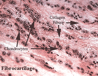
FIBROCARTILAGE
|
Fibrocartilage is very tough, made up of thickly interlaced collagen fibers. It's often found near joints and has the unique ability to merge with tendons or other neighboring tissues.
Fibrocartilage can be found near the edges of articular cavities and in the front area of the pelvic girdle. It is the primary component of the discs between your vertebrae. Fibrocartilage absorbs shocks to prevent damage to the bones, enhances the sturdiness of joints while still allowing movement and deepens joint sockets to reduce the chances of dislocation. |
|
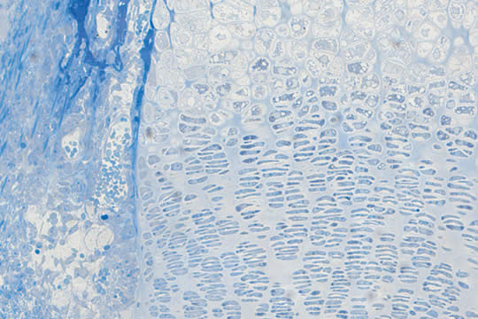
2. BONE
|
Bone is a very hard and rigid tissue with protein ossein present in the matrix. It has white collagenous fibers that interlaces with each other and has is heavily deposited with salts of calcium and phosphorus. The organic and inorganic matter in the matrix form about 30% and 70% respectively.
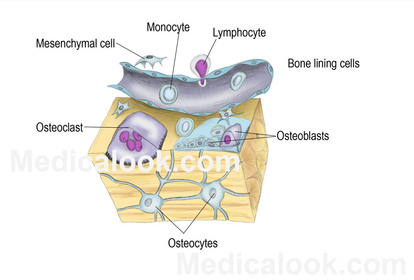
The bone tissue is made up of three types of cells - Osteocytes-These are the inactive bone cells which have numerous processes and thus has a stellate appearance. Osteoblasts- These cells form new bone cells and are present in the periosteum and the endosteum. They add cells to the lamellae by dividing. The new bone cells are converted to osteocytes when the lamellae gets deposited with inorganic salts. Osteoclasts- These cells are called as bone phagocytes. They are large, multinucleated cells present on the surface of the spongy bone. These cells when activated can digest small portions of spongy bone and release calcium into the blood stream. The external surface of the bone is covered by a membrane called periosteum. It has an outer fibrous and inner cellular layer. This is richly supplied by blood vessels and has odontoblast cells. Long bones have a hollow cavity in the center filled with bone marrow. This cavity is lined by a membrane called the endosteum. Similar to the periosteum there are odontoblast cells present in the endosteum also. The odontoblast cells are responsible for producing bone cells that result in the bidirectional growth of the bone. |
|
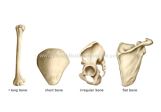
TYPES OF BONES
STRUCTURE OF BONE (Transverse section)
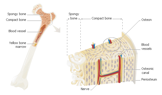
The bone matter can be divided into periosteum, matrix, endosteum and bone marrow.
Periosteum is the outer membrane of the bone whereas the endosteum is the lining of the marrow cavity that are normally seen in long bones.
The matrix is hard, dense and formed of protein called ossein. Structurally the bone has two parts-
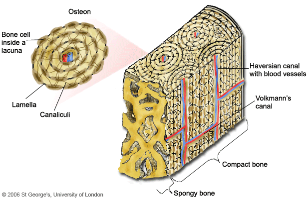
Compact bone- It forms the dense outer region of all bones and is of uniform texture without any spaces. It shows the presence of numerous concentric arrangements, the Haversian system.
Spongy bone- They are found in the deeper parts of the long bones. This does not have any Haversian systems. This part shows fine irregular bony plates known as trabaculae. The trabaculae has wide spaces which are filled in by bone marrow.
Periosteum is the outer membrane of the bone whereas the endosteum is the lining of the marrow cavity that are normally seen in long bones.
The matrix is hard, dense and formed of protein called ossein. Structurally the bone has two parts-
Compact bone- It forms the dense outer region of all bones and is of uniform texture without any spaces. It shows the presence of numerous concentric arrangements, the Haversian system.
Spongy bone- They are found in the deeper parts of the long bones. This does not have any Haversian systems. This part shows fine irregular bony plates known as trabaculae. The trabaculae has wide spaces which are filled in by bone marrow.
|
|
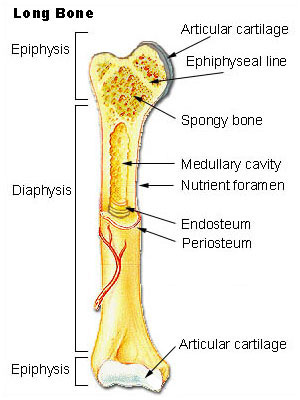
Longitudinal section of long bone
|
Long bones consists of a diaphysis and an epiphysis
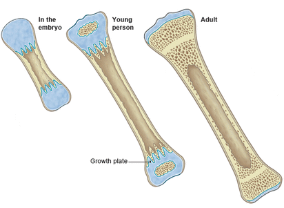
Diaphysis is the tubular shaft that forms the axis of long bones. It is composed of compact bone that surrounds the medullary cavity. Epiphyses is the expanded ends of long bones. The exterior is compact bone, and the interior is spongy bone. The joint surface is covered with articular (hyaline) cartilage. Periosteum is the double-layered protective membrane. The outer fibrous layer is dense regular connective tissue and inner osteogenic layer is composed of osteoblasts and osteoclasts, richly supplied with nerve fibers, blood, and lymphatic vessels. Endosteum is a delicate membrane covering internal surfaces of bone cavity. Marrow cavity- the shaft of the long bones is hollow enclosing a cavity called the marrow cavity. This cavity is filled with with a soft fatty tissue called the bone marrow. Bone marrow is of two type- Red bone marrow- present in the spongy parts of the long bone (epiphysis). It is highly vascular and red due to the presence of iron. It gives rise to different types of blood cells. Yellow bone marrow- present in the shaft of the long bones (diaphysis). It is composed of fatty tissue and stores fat. This marrow produces blood cells at the time of excessive blood loss. |
|
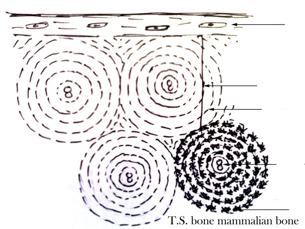
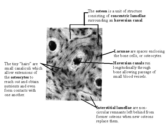
Haversian system
|
A Haversian system, is a series of concentric circles, called lamellae. It is also called as osteon and is found in the compact bone of humans. The middle of each Haversian system is present a hollow tube that holds a blood vessel. The Haversian systems line up next to each other creating a dense structure, each bone thus has many Haversian systems.
The lamellae of each Haversian system shows a series of spaces called lacunae. The osteocytes are present in these lacunae. The osteocytes, along with the collagen and calcium phosphate make up the matrix of the lamellae. This makes the compact bone a very strong structure. The osteoblasts after the deposition of calcium and phosphorous in the lamellae become osteocytes. Nutrition is provided to the bone tissue by the blood vessels running down the middle of the Haversian canal, the canal also has nerves and lymph vessels. There are some lamellae present between the Haversian system that is not concentric, these are called intestitial lamellae. Some lamellae lie parallel to the circumference of the bone and are called circumferential lamellae. Significance of Haversian system: The canaliculi connects the lacunae with each other and with the Haversian canal. Blood vessels and in the Haversian canal bring oxygen and nutrients. The haversian system thus helps in distribution of these to the osteocytes in the lacunae. |
|
Difference between bone and cartilage
Bone
|
Cartilage
|
THE SKELETAL SYSTEM (chapter 8)
|
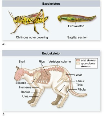
The hard parts of the animal body forms the skeletal system or skeleton.
Skeleton is of two types: Exoskeleton- hard parts on the surface of the body. Endoskeleton –hard parts present inside the body. Parts of the skeletal system
|
|
Functions of skeleton
|
|
Divisions of the skeletal system
AXIAL SKELETON
|
Axial skeleton forms the longitudinal central axis of the body. It includes the
|
|
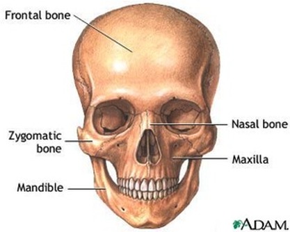
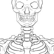
SKULL
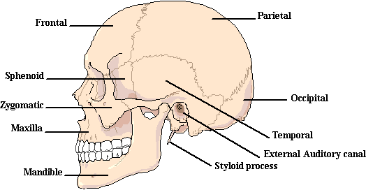
Skull includes the
|
|
|
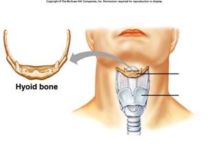
Hyoid bone is the only bone in the body that does not articulate with any other bone.
|
|
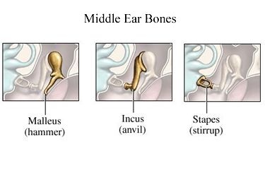
The middle ear has three small bones-
The strirrup is the smallest bone in the body. |
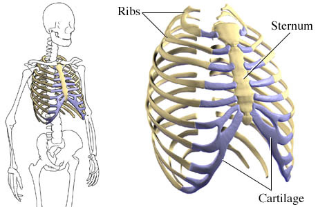
THE RIBCAGE- RIBS AND THE STERNUM
|
The ribcage has twelve pairs of ribs, that articulate with the vertebral column. They are flat, narrow bones with a distinct bow-shaped curve.
The first seven ribs are called true ribs and are attached to the sternum by bars of hyaline cartilage. The cartilages of the 8th, 9th and 10th ribs are joined to the cartilage of the cartilage of the 7th rib. These three pairs of ribs are known as vertebrochondral ribs. The last two pairs of ribs have free ends which are not attached to the sternum at all. They are floating ribs. The vertebrochondral ribs and the floating ribs are collectively known as false ribs. The sternum is a long, flat, dagger-shaped bone. It is about 15 - 18 cm long and is found in the center of the chest region. The first seven pairs of ribs are attached to the articulating facets on the sides of the sternum. |
VERTEBRAL COLUMN
|
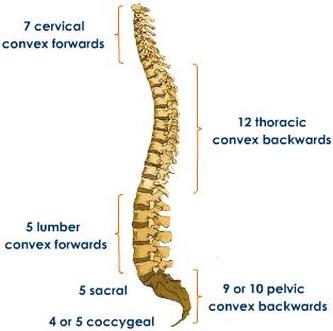
The vertebral column is the central part of the skeleton, that supports the skull and serves as attachment for the ribs, the pectoral and pelvic girdles. It encloses the neural cavity that houses the spinal cord.
The vertebral column is formed of separate bones, the vertebrae. These are arranged one above the other. Since, these vertebrae are attached to each other by a fibrous cartilaginous discs they form a flexible column. Each vertebra has articular surfaces above and below, which allow articulation movement between them. The vertebral column consists of 33 vertebrae. There are five regions according to their position and structure. The five regions consist of: Seven cervical (neck) vertebrae, Twelve thoracic (chest) vertebrae, Five lumbar vertebrae, Five fused sacral vertebrae, and Four fused vertebrae. |
|
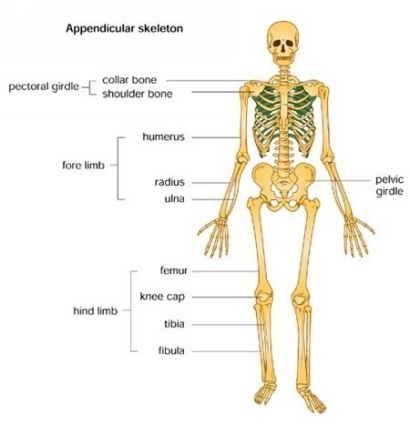
APPENDICULAR SKELETON
|
It is the skeleton that forms the appendages of the body.
It comprises of
|
|
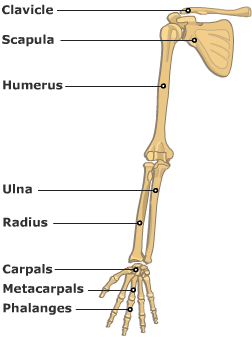
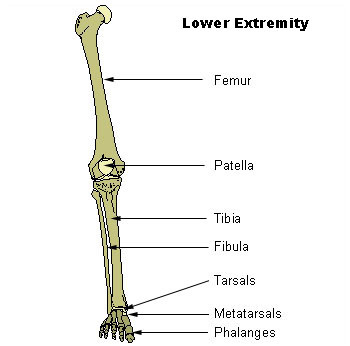
BONES OF UPPER AND LOWER LIMBS
|
Bones of upper limb-
Bones of lower limb
|
Bones of the arms and legs
|
|
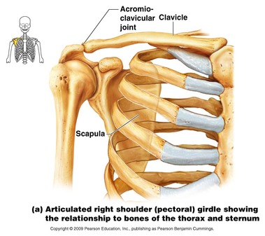
GIRDLES
|
PECTORAL GIRDLE
The bones of the girdle are-
The humerus of the upper limb fits into a cavity on the pectoral girdle called as the glenoid cavity. |
|
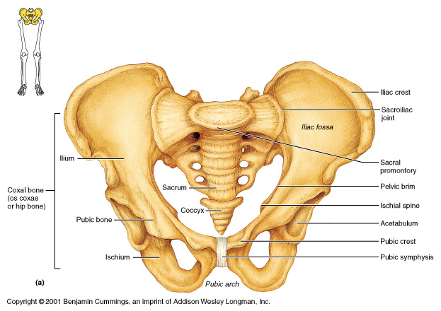
PELVIC GIRDLE
The bones of the girdle are-
The head of the femur fits into a cavity in the pelvic girdle called as the acetabulum or the acetabular cavity. |
|
JOINTS
|
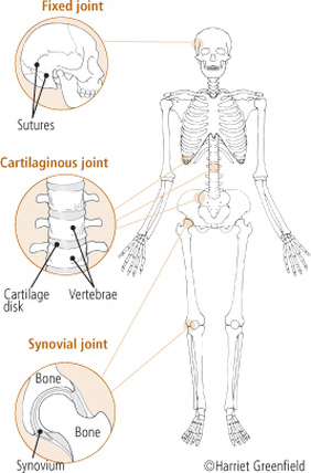
The bones articulate at different places to facilitate the movement. Joints are classified as-
Fibrous joints are held together by only a ligament. These are also called as synarthrosis. Examples are where the teeth are held to their bony sockets and at both the radio-ulnar and tibio-fibular joints. Cartilagenous joints occur where there are connections between the articulating bones and this is made up of cartilage. These are also called amphiarthrosis. The example of this is joint between vertebrae in the spine. |
|
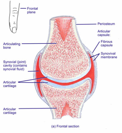
SYNOVIAL JOINT
|
These are called perfect joints or diarthrosis or freely movable joints.
A synovial joint is one in which the ends of the bones are enclosed in a capsule containing a thick, slippery liquid called synovial fluid. The capsule is made of strong, fibrous tissue and is lined with a membrane called the synovial membrane. The bone ends are covered in a smooth layer of a tough, rubbery substance known as cartilage. Synovial Fluid The synovial fluid in the joint capsule has four important functions:
|
TYPES OF FREELY MOVABLE JOINTS
|
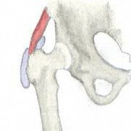
Ball and socket joint:
The ball like end or the head of one bone fits into a socket or concavity of the other. The bone with the ball like end can be freely rotated. These types of joints are - The shoulder joint where the head of the humerus bones articulates in the glenoid cavity of the pectoral girdle. The hip joint the head of the femur fits into the acetabular cavity of the pelvis girdle. |
|
|
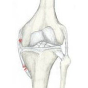
Hinge joint:
In this type of joint the protruberance of one bone fits into the depression of another. Here the bone with the protruberance can be swung in one direction. The movement is like opening and closing of a door. The examples of this type of joint are- Knee and elbow joints and between the phalanges of digits. |
|
|
Pivot joint:
In this type of joint one of the bones is fixed and has a projection, this fits into the concavity of the other and can be freely rotated. It is seen between the atlas and the axis such that the skull freely rotates on the odontoid process of the second vertebra. |
|
|
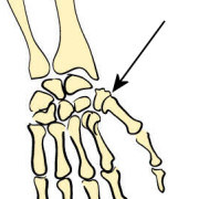
Saddle joint:
These are similar to the ball and socket, however the bones are not properly developed hence there is no free movement. The joint between the metacarpal and the carpal of the thumb. |
|
|
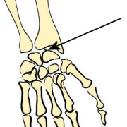
Condyloid or gliding joints
In this type of joints the articulating bones can slide upon one another. However the movements are restricted. The joints between the vertebre and between the radio- ulna and the carpals are examples of such joints. |
|
| bqlocomotion.doc |
