ISC 12> APPLICATIONS OF BIOLOGY>6. BIOMEDICAL ENGG.
SCOPE OF SYLLABUSBiomedical Engineering: (only basic concepts)
Instruments – ECG, EEG, CT scan, ultrasound, MRI, pacemakers, implants, disposables, external prosthesis. Students should know the instruments used for diagnosis of various disorders. Details are not required. |
CLASS PRESENTATION |
WHAT IS BIOMEDICAL ENGINEERING?
Biomedical Engineering (BME) is the application of engineering principles and techniques to the medical field. It combines the design and problem solving skills of engineering with medical and biological sciences to help improve patient health care and the quality of life of individuals.
|
|
Recent advances in medical technology have resulted in development of a number of instruments and devices that have revolutionised the medical world. Earlier medical practitioners had only the traditional instruments to diagnose and monitor diseases. With the advance in technology hospitals and medical centers are equipped with various advanced and sophisticated instruments.
Instruments are tools used for a particular work. These instruments can be grouped as- Diagnostic instruments- These are instruments used for diagnosis. Eg. ECG Imaging instruments- These produce diagnostic images. Eg. CTScan Therapeutic instruments- These are used for curing a disease. Eg. Pace makers |
DIAGNOSTIC INSTRUMENTS
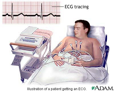
Electrocardiograph(ECG)
This is a sensitive instrument that records electric impulses produced by cardiac muscles of different heart chambers during every cardiac cycle.
The technique of recording the electrical voltages produced by heart muscles during one cardiac cycle is called electrocardiography.
Wilhelm Einthoven is considered father of electrocardiography.
Two electrodes are placed on the chest above the heart and the third is a reference connection to the limbs.
The signal (of only a few millivolts) is amplified before being displayed on a CRT oscilloscope screen or recorded on a sensitive chart recorder.
Electrocardiogram is the graphic record of the spread of cardiac impulses through heart chambers.
It gives information about the rate and rhythm of the heart and the condition of the heart muscles.
It helps to diagnose heart diseases like:
(i) Myocardial infarction.
(ii) Deviation from normal pattern of heart beat.
(iii) Coronary heart diseases etc.
|
|
http://video.about.com/heartdisease/Electrocardiogram.htm
|
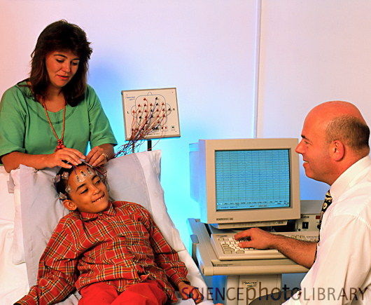
Electroencephalograph (EEG)
An instrument that records spontaneous electrical activity of the brain when electrodes are placed on the head scalp.
The graphic recording is called electroencephalogram.
Hans Berger recorded the first EEG
Recording of the electrical activity of the brain with the help of surface electrodes placed on the scalp is called electroencephalography
Since normal EEG tracing show certain characteristic feature in amplitude and frequency, deviation caused can be detected in terms of changes in the variables, as follows
— Low frequency alpha waves (8—13 Hz) are produced, when the brain is in a relaxed state.
— Theta waves occur during emotional stress.
— Delta waves occur during deep sleep.
— High frequency beta waves are associated with mental activity in the frontal lobe and during periods of sensory stimulations.
ECG is used to detect abnormal brain activity associated with
(i) tumours
(ii) trauma
(iii) epilepsy
(iv) hematomas and
(v) disorders of unconsciousness and confusion.
(vi) degeneration of brain tissue (dementia)
It can be used to identify ‘brain death’.
|
|
|
COMPETITIVE FOCUS
OTHER DIAGNOSTIC INSTRUMENTS-SQUID- (SUPER CONDUCTING QUANTUM INTERFERENCE DEVICE) – It helps in the study of weaker magnetic fields of brain.
Another new technology, magnetoencephalographly (MET) is useful in the study of brain functioning and finding the causes of brain diseases.
When electrodes are applied directly to the brain, during surgery such a record is called electrocorticogram.
Echoencephalogram- when ultrasonic sound waves are used.
Electromyography (EMG)- graphic recording of muscle action potentials. This helps in diagnosing neuro-muscular or muscular diseases.
Autoanalyser- An automatic computer controlled device, used to estimate various biochemical substances like glucose, cholesterol, urea etc present in body fluids.
Endoscopy- A technique that can directly peep into the human body by using an endoscope. It can view hollow organs like food pipe, wind pipe, bladder, intestines and uterus.
IMAGING INSTRUMENTS
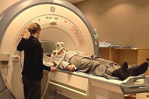
MRI- Magnetic Resonance imaging- or NMR- Nuclear magnetic resonance imaging
It is a tool used for obtaining images of any organ or tissue in any plane.
It was developed by Bloch and Purcell. Its medical use was reported by Raymond Damadian.
It is based on magnetic resonance generated by nuclei of hydrogen atoms when exposed to an external magnetic field.
It is used for –
It was developed by Bloch and Purcell. Its medical use was reported by Raymond Damadian.
It is based on magnetic resonance generated by nuclei of hydrogen atoms when exposed to an external magnetic field.
It is used for –
- Detection of tiny lesions and multiple sclerosis on brain and spinal cord.
- Examination of cancerous bone, pelvic organs, urinary bladder, joints etc.
- Identification of Parkinson’s disease.
- Measuring blood flow and assess heart disease and stroke.
- Study of joints and musclo-skeletal disorders.
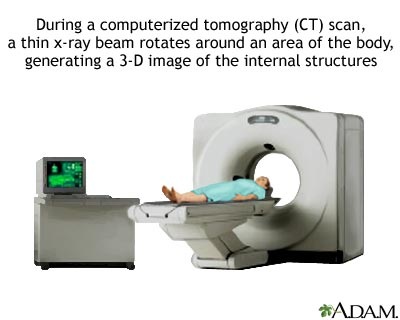
Computerized or Computed Tomography (CT) /computerized axial tomographic scanning (CAT scan)
It is a special type of radiograph which combines X-ray imaging with computer technique permits visualization of internal organs and body structures with greater definition and clarity.
It was developed in 1972 and Godfrey Hounsefield along with Allen Cormack was awarded Nobel Prize in 1978.
CT uses a movable X-ray tube that is rotated around the patient’s body, i.e. any organ.
Opposite to the tube (on the other side of the organ) are X-ray detectors, which record the rays coming out of the body.
Uses of CT Scan
CT is useful for diagnosis of diseases of brain, spine, chest and abdomen. It can also detect blood clots, cysts and tumors.
It is also useful in detecting tumours, and their spread to nearby organs.
It helps to determine the feasibility of operative treatment and to asses the results of treatment.
Before CT technique the doctors had to perform exploratory operations by inserting tubes, cameras, probes etc. into the patients body. This was risky interms of the patients and time consuming for the doctors.
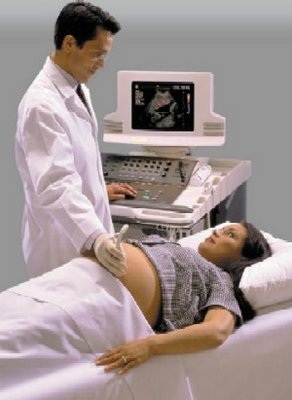
Ultrasound imaging or Sonography
|
Ultrasound of frequency between 1-15 MHz is beamed into the body and the returning echoes are detected.
The ultrasound waves pass unimpeded through a homogenous tissue. But when they meet another tissue or organ of a different density a partial reflection takes place and the coefficient of reflection depends on the difference in densities of the two tissues. Uses — It is used to assess foetal growth — It provides pictures of blood flow based on Doppler effect. — It is useful for detecting diseases of heart, kidney, gall bladder, etc. |
|
|
|
|
THERAPEUTIC INSTRUMENTS
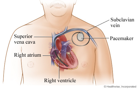
PACEMAKERS
A pacemaker is a small device that's placed in the chest or abdomen to help control abnormal heart rhythms. This device uses electrical pulses to prompt the heart to beat at a normal rate. It is a life saving electronic pulse generator implanted near heart to enhance and control normal heart beat.
A fully implantable pacemaker was used by Chardack
It consists of a pulse generator and an electrode. The entire assembly is implanted under the skin below the collar bone.A pacemaker can relieve some arrhythmia symptoms, such as fatigue and fainting. A pacemaker also can help a person who has abnormal heart rhythms resume a more active lifestyle.
|
|
|
COMPETITIVE FOCUS
Other therapeutic instruments include-
Medical lasers- High energy particles of light amplified by stimulated emission or radiation. Eg:- Argon, Neon and carbon dioxide lasers. Can be specifically targeted to any kind of tissue as powerful energy beams. Used in many kinds of surgical operations eg:- in treatment of tumours especially of retina and brain; the cancer cells (tumour) are localised, laser beam is focussed on it, the cells are selectively burnt out without affecting normal cells. Laser photobleaching gives details of hormone action mechanism.
Intra aortic balloon pump- It is used in cases of heart failure, involving inefficient pumping action of the cardiac ventricle. It consists of a special balloon positioned in the descending thoracic aorta. The pumping action is brought about by connecting it through a tube to an external pumping machine. It can be used for long time till the patient becomes normal.
MEDICAL DEVICES
Implants
An implant is a tissue or organ inserted in the body to replace the defective or diseased tissue or organ.
These implants are non-toxic and biocompatible.
It includes:
Artificial heart valves, vascular grafts, Coronary Artery Bypass Grafting (CABG) and transplantation.
Transplantation is the replacement of injured or diseased tissue.
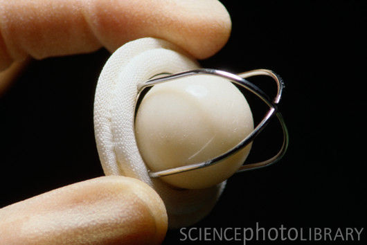
Artificial heart valves are used for replacing defective heart valves. These are of two types mechanical and tissue valves.
|
|
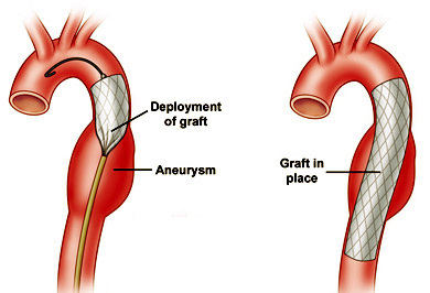
Vascular grafts or arterial grafts are used for reconstruction of defective arteries due to atherosclerosis or aneurysm.
|
|
|
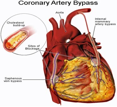
CABG (CORONARY ARTERY BYPASS GRAFT) is used to increase blood supply to the heart by bypassing the blocked region of the coronary artery.
|
|
|
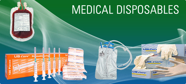
Disposables
These are medical devices which are used externally only for once and then discarded. These include- syringes, urinary catheter, oxygenator, blood bag and blood dialyser. |
|
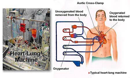
Oxygenators
|
The oxygenator is main element of the heart-lung machine, which takes over the work of the lungs (adding oxygen to and removing carbon dioxide from the blood).
Inside the oxygenator, blood is gently channelled along capillary membranes. The inner lumen of the fibres is streamed with oxygen or oxygen enriched air. Oxygen diffuses through the microporous membrane into the blood, while carbon dioxide diffuses out of the blood into the gas stream and is thereby removed. Most oxygenators provide a heat exchanger in order to maintain the correct temperature of the patient’s blood. The oxygenated blood is channelled back to the patient. |
|
|
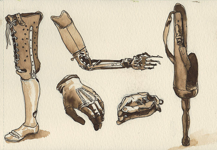
Prosthesis
It refers to the implantation of an artificial substitute for a body part. Myoelectrical artificial arm uses muscle impulse and patient is able control the movement of prosthetic wrist and hand. It can function like a real hand. Generally the term prosthetic is used to describe external attachments and appendages. Examples of prosthetics in everyday life are glasses, contact lenses, replacement legs and arms, and anything else that augments the body and extends the use of a body part. External prosthesis or replacement is exemplified by the denture and Jaipur foot. Dr. P.K.Sethi has developed artificial limb resembling the natural foot. Internal prosthesis includes intra-ocular lens, cardiac pacemaker, heart-lung machine etc. |
|
|
| ||||
