ISC 11> NERVOUS SYSTEM> PERIPHERAL NERVOUS SYSTEM
PERIPHERAL NERVOUS SYSTEM
Peripheral nervous system consists of- cranial nerves arising from the brain and the spinal nerves arising from the spinal cord.
|
Sensory and motor nerve fibers.
|
|
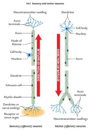
AFFERENT (SENSORY) NERVES
They conduct sensory impulses from receptors in the peripheral tissue to the CNS. The cell bodies of these fibers are called afferent neurons. |
EFFERENT (MOTOR) NERVES
They conduct motor impulses from central nervous system to the effectors. The cell bodies of these fibers are called efferent neurons. |
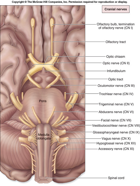
CRANIAL NERVES
|
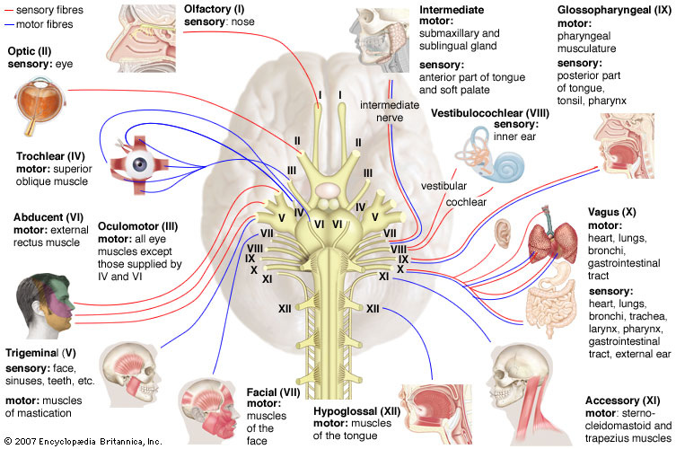
The cranial nerves are 12 pairs of nerves that can be seen on the ventral (bottom) surface of the brain. Some of these nerves bring information from the sense organs to the brain; other cranial nerves control muscles; other cranial nerves are connected to glands or internal organs such as the heart and lungs.
|
| CRANIAL NERVES | |||
|---|---|---|---|
| Number | Name | Function | TYPE |
| I | Olfactory Nerve | Smell | Sensory |
| II | Optic Nerve | Vision | Sensory |
| III | Oculomotor Nerve | Motor control of eyeball and some eye muscles and eye lids | Motor |
| IV | Trochlear Nerve | Precise movement of the eye for visual tracking or fixation on an object | Motor |
| V | Trigeminal Nerve | Somatosensory information (touch, pain) from the face and head; muscles for chewing. | Mixed |
| VI | Abducens Nerve | Eye movement- control of eye muscles | Motor |
| VII | Facial Nerve | Taste (anterior 2/3 of tongue); somatosensory information from ear; controls muscles used in facial expression. | Mixed |
| VIII | Vestibulocochlear Nerve | Hearing; balance | Sensory |
| IX | Glossopharyngeal Nerve | Taste (posterior 1/3 of tongue); Somatosensory information from tongue, tonsil, pharynx; controls some muscles used in swallowing. | Mixed |
| X | Vagus Nerve | Sensory, motor and autonomic functions of viscera (glands, digestion, heart rate) | Mixed |
| XI | Spinal Accessory Nerve | Controls muscles used in head movement. | Motor |
| XII | Hypoglossal Nerve | Controls muscles of tongue | motor |
SPINAL NERVES
|
There are a total of 31 bilaterally-paired spinal nerves :
There are networks of intersecting nerves 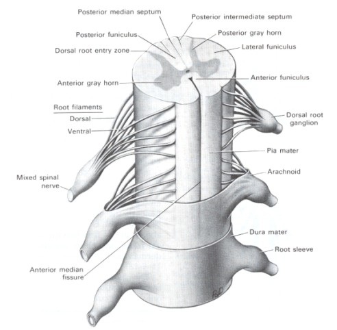
|
|
Spinal Nerves
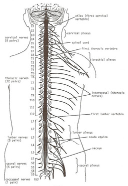
There are 31 pairs of spinal nerves that leave the spinal cord and pass through the intervertebral foramina in the vertebral column. The spinal nerves are named according to the regions of the vertebral column with which they are associated: 8 cervical, 12 thoracic, 5 lumbar, 5 sacral, and 1 coccygeal. Note that there are 8 cervical nerves and only 7 cervical vertebrae and that there are 1 coccygeal nerve and 4 coccygeal vertebrae.
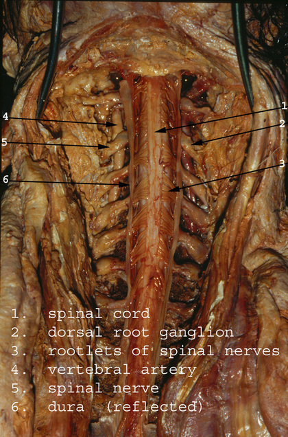
Each spinal nerve is connected to the spinal cord by two roots: the anterior root and the posterior root. The anterior root consists of bundles of nerve fibers carrying nerve impulses away from the central nervous system. Such nerve fibers are called efferent fibers. Those efferent fibers that go to skeletal muscle and cause them to contract are called motor fibers. Their cells of origin lie in the anterior gray horn of the spinal cord.
The posterior root consists of bundles of nerve fibers that carry impulses to the central nervous system and are called afferent fibers. Since these fibers are concerned with conveying information about sensations of touch, pain, temperature, and vibrations, they are called sensory fibers. The cell bodies of these nerve fibers are situated in a swelling on the posterior root called the posterior root ganglion.
At each intervertebral foramen the anterior and posterior roots units to form a spinal nerve
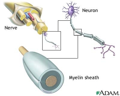
NERVE FIBER
Nerve fibers are axons of a neuron.
PROPERTIES OF NERVE FIBER
EXCITABILITY-
It is the property of the cells to respond to stimuli from the external or internal environment.
CONDUCTIVITY-
Conductance of nerve impulse means, spread of wave of depolarization.
It is the property of the cells to respond to stimuli from the external or internal environment.
CONDUCTIVITY-
Conductance of nerve impulse means, spread of wave of depolarization.
NERVE IMPULSE CONDUCTION
Nerve impulse is an electric current that travels along the axons due to ions moving through voltage-gated channels in the neurone.
Neurons can respond to stimuli and conduct impulses because a membrane potential is established across the cell membrane. There is an unequal distribution of ions (charged atoms) on the two sides of a nerve cell membrane.
If an electrode placed inside a neuron and the other one outside, the voltmeter will measure the difference in the distribution of ions on the inside versus the outside. This will be approximately -70 mV (mV = millivolts).
Thus it can be said that the inside of the neuron is slightly negative relative to the outside. This difference is referred to as the Resting Membrane Potential. The membrane is said to be polarised.
This potential is maintained by two ions: sodium (Na+) and potassium (K+).
There is an unequal distribution of these two ions on the two sides of a nerve cell membrane because of the sodium potassium pump. There is a higher concentration of sodium on the outside than the inside and a higher concentration of potassium on the inside than the outside.
The nerve cell membrane also contains special passageways for these two ions that are commonly referred to as gates or channels.
Thus, there are sodium channels and potassium channels. These channels are the only way that these ions can diffuse through a nerve cell membrane.
IN A RESTING NERVE CELL MEMBRANE, all the sodium channels are closed and some of the potassium channels are open. Thus the sodium cannot diffuse through the membrane & largely remains outside the membrane. However, some potassium ions are able to diffuse out.
Therefore, there are lots of positively charged potassium ions just inside the membrane and lots of positively charged sodium ions and some potassium ions on the outside. THIS MEANS THAT OVERALL THERE ARE MORE POSITIVE CHARGES ON THE OUTSIDE THAN ON THE INSIDE. This potential will be maintained until the membrane is disturbed or stimulated.
An action potential is a very rapid change in membrane potential that occurs when a nerve cell membrane is stimulated. Specifically, the membrane potential goes from the resting potential (typically -70 mV) to some positive value (typically about +30 mV) in a very short period of time (just a few milliseconds).
The Action potential-
A stimulus causes the sodium gates (or channels) to open and, because there's more sodium on the outside than the inside of the membrane, sodium diffuses rapidly into the nerve cell. All these positively-charged sodiums rushing in causes the inside of the membrane potential to become positive. This is called as action potential. The membrane is now said to be deploarised. These sodium channels open only briefly, then close again.
The potassium channels then open, and, because there is more potassium inside the membrane than outside, positively-charged potassium ions diffuse out. As these positive ions go out, the inside of the membrane once again becomes negative with respect to the outside. This is repolarisation.
Action potentials occur only when the membrane in stimulated (depolarized) enough so that sodium channels open completely. The minimum stimulus needed to achieve an action potential is called the threshold stimulus.
All-or-None Law - action potentials occur maximally or not at all. In other words, there's no such thing as a partial or weak action potential. Either the threshold potential is reached and an action potential occurs, or it isn't reached and no action potential occurs.
Impulse conduction- an impulse is the movement of action potentials along a nerve cell. Action potentials are localized. So, when one occurs, only a small area of membrane depolarizes. As a result, for a split second, areas of membrane adjacent to each other have opposite charges. An electrical circuit develops between these oppositely-charged areas. This stimulates the adjacent area and, therefore, an action potential occurs. This process repeats itself and action potentials move down the nerve cell membrane. This 'movement' of action potentials is called an impulse.
NERVE IMPULSE CONDUCTION
Neurons can respond to stimuli and conduct impulses because a membrane potential is established across the cell membrane. There is an unequal distribution of ions (charged atoms) on the two sides of a nerve cell membrane.
If an electrode placed inside a neuron and the other one outside, the voltmeter will measure the difference in the distribution of ions on the inside versus the outside. This will be approximately -70 mV (mV = milliv
If an electrode placed inside a neuron and the other one outside, the voltmeter will measure the difference in the distribution of ions on the inside versus the outside. This will be approximately -70 mV (mV = milliv
|
|
In an unmyelinated axon, the action potential is propagated along the entire membrane. The action potential jumps along the fiber as it is regenerated at each node. This is called saltatory conduction.
|
|
IMPULSE CONDUCTION IN MYELINATED NERVE FIBRES
1 Velocity of impulse conduction is quite high (up to 120 m/sec). 2 Action potential develops only in areas of nodes of Ranvier. 3 Impulse conduction is saltatory. 4 Less energy is spent. |
IMPULSE CONDUCTION IN NONMYELINATED NERVE FIBRES
1 It is slow (up to 10-20 m/sec). 2 It develops throughout the nerve fibre. 3 Impulse conduction is continuous. 4 More energy is spent. |
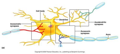
SYNAPSE
Synapses are specialized junctions through which cells of the nervous system signal to one another and to non-neuronal cells such as muscles or glands.
|
|
At a chemical synapse, one neuron releases neurotransmitter molecules into a small space (the synaptic cleft) that is adjacent to another neuron. These molecules then bind to the neuroreceptors on the receiving cell's side of the synaptic cleft. Finally, the neurotransmitters must be cleared out of the synapse efficiently so that the synapse can be ready to function again as soon as possible.
|
TYPES OF SYNAPSE
1) Synapses on cell body are often inhibitory, i.e. axosomatic 2) Synapses on dendritic spines are often excitatory, i.e. axodendritic 3) Synapses on axon terminals are often modulatory, i.e. axoaxonic controlling the amount of transmitter released Synapse |
|
NEUROTRANSMITTERS
These are chemicals secreted by a presynaptic neuron that transmit impulse to the next neuron. The neurons that produce acetylcholine is are called cholinergic and those producing nor epinephrine are called adrenergic.
The enzyme acetylcholinesterase splits acetycholine into acetic acid and choline.
The enzyme monoamine oxidase inactivates norepinephrine.
GABA- Gaba Amino-butryic acid is an inhibitory neurotransmitter.
Dopamine, Histamine, Serotonin are some examples of neurotransmitters.
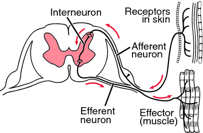
REFLEX ACTION
|
|
|
REFLEX ACTION
Spontaneous automatic, involuntary, nerve mediated activity produced at the unconscious level by stimulating specific receptors.
Spontaneous automatic, involuntary, nerve mediated activity produced at the unconscious level by stimulating specific receptors.
|
The path traveled by an impulse in a reflex action is called as reflex arc.
COMPONENTS AND FUNCTIONS OF THE REFLEX ARC:
|
|
TYPES OF REFLEX
|
UNCONDITIONED REFLEX
1 It is an innate characteristic of an animal. 2 It does not require previous learning or experience. 3 It is shown by all individuals. 4 It is a response to a stimulus which normally evokes that response. 5 It is not lost even if the stimulus is not received for a long time. It cannot change. 6 It does not need additional receptor and cerebral centre. |
CONDITIONED REFLEX
1 It is an acquired behaviour of an animal. 2 It gradually develops by training (repetition of a definite stimulus). 3 It is shown only by individuals trained for it. 4 It is a response to a stimulus other than the one which normally evoles that response. 5 It is lost, if the conditioned stimulus is discontinued for some time. It can change also. 6 It needs additional receptor and cerebral centre. |
AUTONOMIC NERVOUS SYSTEM
|
Sympathetic System
1. Sympathetic fibres originate from cell bodies in the grey matter of spinal cord. 2. Neurons of sympathetic fibres are situated in the sympathetic ganglia that form a chain along the bentrolateral sides of vertebral column. 3. Preganglionic fibres of sympathetic neurons are short. 4. Postganglionic fibres of sympathetic neurons are long. 5. Neurotransmitter released in the effector is adrenaline or epinephrine and noradrenaline or norepinephrine. 6. Sympathetic fibres are called adrenergic. 7. Sympathetic system accelerates activities and prepares the body against adverse condition for fight or flight. |
Parasympathetic System
1. Parasympathetic fibres arise from cell bodies present in the brain and grey matter of spinal cord in the sacral region. 2. Neurons of parasympathetic fibres remain isolated. 3. Preganglionic fibres of parasympathetic fibres are long. 4. Postganglionic fibres are short. 5. Neurotransmitter released in the effector is acetylcholine. 6. Parasympathetic fibres are called cholinergic. 7. Parasympathetic system restores normalcy and provides feeling of relaxation, comfort and pleasure. |
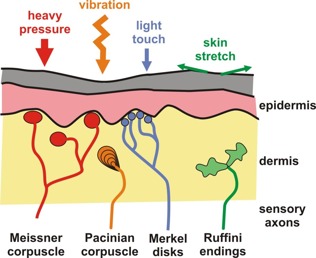
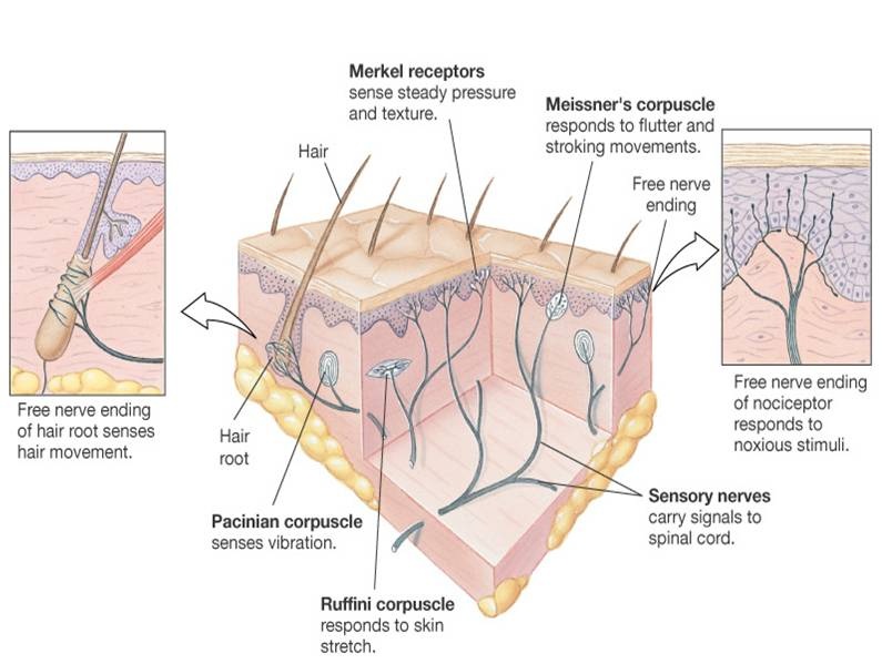
Sensory organs (receptors) monitor the internal and external environment. They transmit signals from the periphery to the CNS for processing. This is critical to maintain homeostasis.
Based on type of stimulus-
Based on type of stimulus-
- Mechanoreceptors respond to physical force such as pressure (touch or blood pressure) and stretch.
- Photoreceptors respond to light.
- Thermoreceptors respond to temperature changes.
- Chemoreceptors respond to dissolved chemicals during sensations of taste and smell and to changes in internal body chemistry such as variations of O2, CO2, or H+ in the blood.
- Nociceptors respond to a variety of stimuli associated with tissue damage. The brain interprets the pain.
|
Sensory organs (receptors) monitor the internal and external environment. They transmit signals from the periphery to the CNS for processing. This is critical to maintain homeostasis.
Based on type of stimulus-
|
|
|
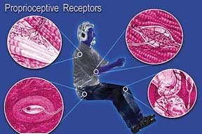
Based on location:
Based on the complexity of structures-
|
|
